Kirjojen hintavertailu – 12 903 724 kirjaa ja 27 kauppaa

Kirjailija

A. Wackenheim

Kirjat ja teokset yhdessä paikassa: 8 kirjaa, julkaisuja vuosilta 1980–2012, suosituimpiin kuuluu Röntgendiagnostik der Wirbelsäule Teil 1 / Roentgendiagnosis of the Vertebral Column Part 1. Vertaile teosten hintoja ja tarkista saatavuus suomalaisista kirjakaupoista.

8 kirjaa

Kirjojen julkaisuvuodet: 1980–2012.

Röntgendiagnostik der Wirbelsäule Teil 1 / Roentgendiagnosis of the Vertebral Column Part 1

Röntgendiagnostik der Wirbelsäule Teil 1 / Roentgendiagnosis of the Vertebral Column Part 1

L. Diethelm; M. Erdélyi; W. Hoeffken; H. Junge; O. Perey; W. Pfeiffer; K. Reinhardt; K. Theiler; G. Töndury; A. Wackenheim; H. Wolfers; W. Zaunbauer

Springer-Verlag Berlin and Heidelberg GmbH Co. K
2011
nidottu
In 1932 there appeared the work of SCHMORL and JUNGHANNS, Die gesunde und kranke Wirbelsiiule im Rontgenbild, which laid the foundations of diagnostic radiology of the spine. Since that time the discipline has been extended and refined in a systematic manner and our knowledge has been greatly enriched by the large number of monographs in which leading experts have assembled additional data. An encyclopedic treatment of all that is currently known requires not only that reference be made to all work published since 1932 in order to reveal the problems that remain but also that a serious attempt be made to contribute to the solution of such problems. For this reason, Professor TONDURY and Professor THEILER of Zurich, who know more than anyone else about the ontogeny of the spine, have been invited to display their erudition in this field. Should contra- dictions or divergent opinions on certain points be revealed through this confrontation with radiologically obtained findings, so much the better; the effect of this could only be to spur workers in both disciplines on to more advanced research. We have only to recall how in Goethe's time the premaxilla was still the subject of controversy, yet this great man pursuing his studies in comparative anatomy and anthropology was able to prove its existence beyond all doubt. Mainz, May 1974 L. DIETHELM Inhaltsverzeichnis - Contents A. Die normale Wirbelsliule ...1 1 I. Phylogenetische Entwicklung des Achsenskelets. Von K. THEILER 1 1. Die Bogenelemente...2. Die Chorda dorsalis ...
Cheirolumbar Dysostosis

Cheirolumbar Dysostosis

A. Wackenheim

Springer-Verlag Berlin and Heidelberg GmbH Co. K
1980
nidottu
It was with great pleasure that I accepted Professor Wackenheim's invitation to write the preface of this most interesting monograph. During the international course on neuroradiology in Obernai, organized by Professor Wackenheim in 1978, an entire day was devoted to discussions about various aspects of stenosis of the lumbar vertebral canal. As Professor Wackenheim and his group, in particular Dr. E. Babin, had thorough· ly studied this field, it was an excellent occasion for the exchange of views through personal contact. Their support of basic views such as attributing the cause of stenosis to a developmental disturbance of growth of the neural element - the vertebral arc- and spontaneous agreement about essentials in classification and nomenclature facili­ tated this exchange. Reading the present monograph made me think back to 1949 when I wrote my first publication on stenosis of the lumbar vertebral canal in a French volume that com­ memorated my teacher in neurosurgery, Professor Clovis Vincent. During the following years it was impossible to publish a more detailed paper on the subject in international journals since their editorial boards did not believe in its occurrence. My first English papers were published in the Journal of Bone and Joint Surgery (1954, 1955), but it was only during the 1970s that this form of stenosis became more universally recog­ nized.
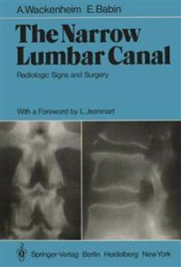
The Narrow Lumbar Canal

The Narrow Lumbar Canal

A. Wackenheim; E. Babin

Springer-Verlag Berlin and Heidelberg GmbH Co. K
2011
nidottu
It is amazing to discover how little importance has been attached to narrow lumbar canal syndromes up to now. Though H. VERBIEST gave a very accurate description in 1949, the neurologist's and neurosurgeon's preoccupations were mainly focused on discal pathology, disregarding the problem of an exclusively bony origin in canalar stenosis. A. WACKENHEIM and E. BABIN have the merit of becoming aware of the impor­ tance and originality of this problem; they organized in the beautiful surround­ ings of the Bischenberg near Strasbourg, a postgraduate course, in which the most eminent European specialists in this field participated. I am very honored to have been asked to write the introduction to this mono­ graphy, which contains all the studies reported and commented on during this meeting. Before considering the problem from the various radiologic points of view, it is in my opinion indispensable to define the term "stenosis." We could not do so more accurately than by assuming the definition proposed by A. WACKENHEIM and E. BABIN and unanimously confirmed by all those who attented the session.
The Veins of the Posterior Fossa

The Veins of the Posterior Fossa

A. Wackenheim; J.P. Braun

Springer-Verlag Berlin and Heidelberg GmbH Co. K
2011
nidottu
It is a privilege and pleasure to write a foreword to this work. Who, twenty years ago, would have thought that a whole book could be devoted to the veins of the posterior fossa? Even today the standard text-books of anatomy only give a paragraph to the subject. Thanks to the dedicated work of rather a few neuroradiologists our knowledge has now reached a very sophisticated level. The careful correlation of anatomical dissection with the angiographic image has achieved this, but without the aid of subtraction (discovered by a world figure in neuroradiology - Professor ZIEDSES DES PLANTES) the subject would not be as advanced as it is. The authors are to be congratulated not only for presenting the most up to date anatomical information but also for showing how wide the applications are in the clinical field by the careful analysis of displacements of veins, many of which were quite unrecognised only a very few years ago. Drs W ACKENHEIM and BRAUN are in the forefront of this discipline to which they and the French school have contributed so much. Also it was their great enthusiasm and drive which led to the birth of the European Society of Neuroradiology, the first meeting of which was held in Colmar in September 1969. The Society has flourished under their guidance and very successful annual meetings have been held for nearly a decade.
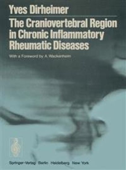
The Craniovertebral Region in Chronic Inflammatory Rheumatic Diseases

The Craniovertebral Region in Chronic Inflammatory Rheumatic Diseases

Yves Dirheimer; A. Wackenheim

Springer-Verlag Berlin and Heidelberg GmbH Co. K
2011
nidottu
Radiology, the youngest of the major medical sciences, has undergone an extraordinary technical evolution since the discovery of X-rays. It began with the development of the different types of tomography and the adoption of many contrast agents, then proceeded rapidly to serioscopy, subtraction of images, direct enlargement, echography, thermography, and xerography. Today, even before all these innovations have come into common use, another branch of radiologic technology has evolved: computerized (axial) tomography. More than just an innovation, its true dimensions are unfore­ seeable. Radiology has become in less than a century an indispensable adjunct to the practice of medicine. The development of radiology as a speciality followed its technical advances, which varied greatly from country to country. This rapid development led quickly to subspecialization, even the very early development of radiotherapy and radiodiagnostics as separate entities. However, the entry of radiology into the university has preserved it a single branch of medicine, avoiding the frequent tendency toward auto­ nomy of the branches of a speciality. Today the fourth generation of radiologists is faced with another deci­ sion: whether to become technologists subjugated to their machinery, to become sub specialists with a single skill, or to remain doctors. The vast majority of this fourth generation has rejected becoming an accessory to a master technique and rather has specialized according to the hippocratic concept of medicine.