Kirjojen hintavertailu – 12 859 819 kirjaa ja 27 kauppaa

Kirjailija

Ameni Jerbi

Kirjat ja teokset yhdessä paikassa: 25 kirjaa, julkaisuja vuosilta 2024–2025, suosituimpiin kuuluu Atipichnye aspekty antinuklearnyh antitel. Vertaile teosten hintoja ja tarkista saatavuus suomalaisista kirjakaupoista.

25 kirjaa

Kirjojen julkaisuvuodet: 2024–2025.

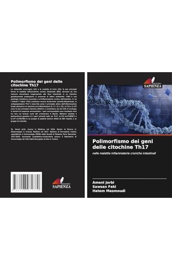
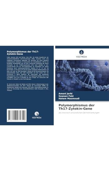
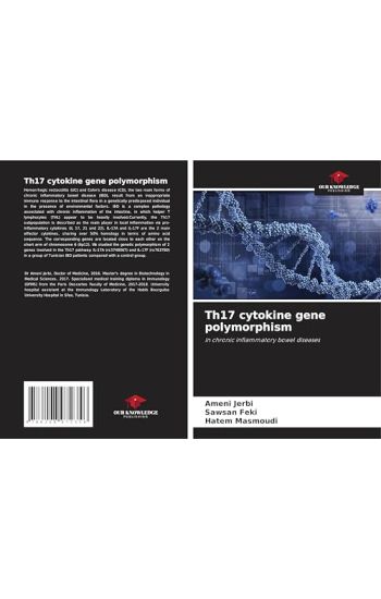
Th17 cytokine gene polymorphism

Th17 cytokine gene polymorphism

Ameni Jerbi; Sawsan Feki; Hatem Masmoudi

Our Knowledge Publishing
2025
nidottu
Hemorrhagic rectocolitis (UC) and Cohn's disease (CD), the two main forms of chronic inflammatory bowel disease (IBD), result from an inappropriate immune response to the intestinal flora in a genetically predisposed individual in the presence of environmental factors. IBD is a complex pathology associated with chronic inflammation of the intestine, in which helper T lymphocytes (THL) appear to be heavily involved. Currently, the Th17 subpopulation is described as the main player in local inflammation via pro-inflammatory cytokines (IL 17, 21 and 22). IL-17A and IL-17F are the 2 main effector cytokines, sharing over 50% homology in terms of amino acid sequence. The corresponding genes are located close to each other on the short arm of chromosome 6 (6p12). We studied the genetic polymorphism of 2 genes involved in the Th17 pathway: IL-17A (rs3748067) and IL-17F (rs763780) in a group of Tunisian IBD patients compared with a control group.
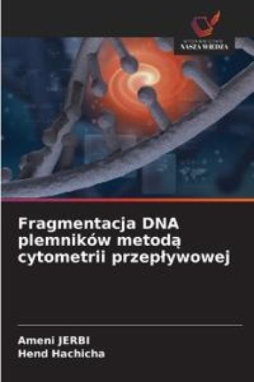
Sperm DNA fragmentation by flow cytometry

Sperm DNA fragmentation by flow cytometry

Ameni Jerbi; Hend Hachicha

Our Knowledge Publishing
2025
nidottu
Sperm DNA fragmentation involves single- or double-strand breaks in the genetic material of sperm cells. It is an anomaly likely to reduce the fertilizing power of spermatozoa. It is therefore one of the causes of male infertility. The study of sperm DNA fragmentation is therefore an integral part of the work-up for male infertility, with spermogram analysis on the one hand, and assisted reproduction techniques on the other. Flow cytometry is one of the approaches used in clinical practice to assess sperm DNA fragmentation. On the other hand, increased sperm DNA fragmentation can be associated with several extrinsic factors (such as heat exposure, smoking, urogenital infections and varicocele) or intrinsic factors related to defective germ cell maturation linked to oxidative stress. Some of these factors may be reversible and/or curable, which could improve sperm DNA quality and the couple's obstetrical prognosis.
Indagine immunologica delle dermatosi bollose autoimmuni
Le dermatosi bollose autoimmuni (ABD) sono malattie autoimmuni specifiche della pelle e delle mucose. Sono caratterizzate dalla presenza di autoanticorpi (auto-Ab) diretti contro molecole di adesione della cute e delle mucose, che determinano un'alterazione della funzione dei loro bersagli antigenici, con conseguente formazione di bolle cliniche e/o erosioni. A seconda del sito di clivaggio, si distingue tra DBAI intraepidermica, caratterizzata da auto-Ac diretti contro le proteine strutturali dei desmosomi che tengono uniti i cheratinociti dell'epidermide, e DBAI subepidermica, caratterizzata dalla presenza di auto-Ac diretti contro i componenti della giunzione dermo-epidermica (DEJ). La diagnosi di DBAI si basa su evidenze cliniche, istopatologiche e immunologiche (rilevamento di auto-Ac circolanti o auto-Ac depositati nei tessuti). L'immunofluorescenza diretta (DIF) una tecnica immunologica eseguita su criosezioni di biopsie peri-lesionali basata sul rilevamento di depositi di Ac e/o frazioni del complemento utilizzando Ac specifici coniugati con fluoresceina.
Investigação imunológica das dermatoses bolhosas auto-imunes
As dermatoses bolhosas auto-imunes (ABD) s o doen as auto-imunes espec ficas da pele e das membranas mucosas. Caracterizam-se pela presen a de auto-anticorpos (auto-Ab) dirigidos contra mol culas de ades o na pele e nas mucosas, levando altera o da fun o dos seus alvos antig nicos, resultando na forma o de bolhas cl nicas e/ou eros es. Dependendo do local de clivagem, feita uma distin o entre DBAI intra-epid rmico, caracterizado por auto-Ac dirigidos contra as prote nas estruturais dos desmossomas que mant m unidos os queratin citos da epiderme; e DBAI subepid rmico, caracterizado pela presen a de auto-Ac dirigidos contra os componentes da jun o dermo-epid rmica (DEJ). O diagn stico da DBAI baseia-se em provas cl nicas, histopatol gicas e imunol gicas (dete o de auto-Ac circulante ou de auto-Ac depositado nos tecidos). A imunofluoresc ncia direta (IFD) uma t cnica imunol gica realizada em criossec es de bi psias peri-lesionais, baseada na dete o de dep sitos de Ac e/ou de frac es do complemento utilizando Ac espec ficos conjugados com fluoresce na.
Immunological investigation of autoimmune bullous dermatoses
Autoimmune bullous dermatoses (ABD) are specific autoimmune diseases of the skin and mucous membranes. They are characterized by the presence of autoantibodies (auto-Ab) directed against skin and mucous membrane adhesion molecules, leading to altered function of their antigenic targets, resulting in clinical bullae and/or erosions. Depending on the site of cleavage, a distinction is made between intra-epidermal DBAI, characterized by auto-Ac directed against the structural proteins of the desmosomes that hold the keratinocytes of the epidermis together; and sub-epidermal DBAI, characterized by the presence of auto-Ac directed against the components of the dermo-epidermal junction (DEJ). The diagnosis of DBAI is based on clinical, histopathological and immunological evidence (detection of circulating or tissue-deposited auto-Ac). Direct immunofluorescence (DIF) is an immunological technique performed on cryosections of peri-lesional biopsies, based on the detection of Ac deposits and/or complement fractions using specific fluorescein-conjugated Ac.
Immunologische Untersuchung von autoimmunen bullösen Dermatosen
Autoimmune bull se Dermatosen (ABD) sind spezifische Autoimmunerkrankungen der Haut und der Schleimh ute. Sie sind durch das Vorhandensein von Autoantik rpern (Auto-AK) gekennzeichnet, die gegen die Adh sionsmolek le der Haut und der Schleimh ute gerichtet sind und die Funktion ihrer antigenen Zielmolek le beeintr chtigen, was klinisch zur Bildung von Blasen und/oder Erosionen f hrt. Je nach Spaltstelle unterscheidet man zwischen intraepidermalen DBAI, bei denen die Auto-AK gegen die Strukturproteine der Desmosomen gerichtet sind, die die Keratinozyten der Epidermis zusammenhalten, und subepidermalen DBAI, bei denen die Auto-AK gegen die Komponenten der dermoepidermalen Junction (DJE) gerichtet sind. Die Diagnose von DBAI beruht auf klinischen, histopathologischen und immunologischen Argumenten (Nachweis von zirkulierenden oder im Gewebe abgelagerten Auto-Ak). Die direkte Immunfluoreszenz (DFI) ist eine immunologische Technik, die an Kryosektionen von peril sionalen Biopsien durchgef hrt wird und auf dem Nachweis von Ak-Ablagerungen und/oder Komplementfraktionen unter Verwendung spezifischer Fluorescein-konjugierter Ak beruht.

Atypische Aspekte von antinukleären Antikörpern
Antinukle re Antik rper (ANA) stellen eine heterogene Gruppe von Autoantik rpern dar, die gegen eine Vielzahl von Antigenen nuklearen oder zytoplasmatischen Ursprungs gerichtet sind. Die Suche nach ANA ist ein h ufiger Anlass in der klinischen Biologie. Das Interesse an ANA liegt im diagnostischen Wert einiger von ihnen f r die Diagnose von Bindegewebserkrankungen und bestimmten organspezifischen oder unspezifischen Autoimmunerkrankungen. Gem dem 1. internationalen Konsens zur Standardisierung der Nomenklatur von AAN "Internationaler Konsens ber Anti-Nuklear-Antik rper-F rbungsmuster" (2014) wird empfohlen, bei der Darstellung von Forschungsergebnissen zu AANs die zytoplasmatische Fluoreszenz und die des mitotischen Apparats mit der Kernfluoreszenz zu verkn pfen . Allerdings mangelt es in der aktuellen Praxis bei der Berichterstattung ber ANA-Ergebnisse an einer Standardisierung zwischen den Labors. Wir haben die verschiedenen Aspekte (nuklear, zytoplasmatisch und mitotisch) der in unserem Labor durchgef hrten ANA gem der ICAP-Klassifizierung analysiert und die klinische Bedeutung der zytoplasmatischen Aspekte untersucht, die mit der Kernmarkierung verbunden sind oder nicht.
Aspectos atípicos dos anticorpos antinucleares

Aspectos atípicos dos anticorpos antinucleares

Ameni Jerbi

Edicoes Nosso Conhecimento
2024
pokkari
Os anticorpos antinucleares (ANA) constituem um grupo heterog neo de autoanticorpos direcionados contra uma ampla variedade de ant genos de origem nuclear ou citoplasm tica. A busca por ANA motivo frequente em biologia cl nica. O interesse nos ANA reside no valor diagn stico de alguns deles para o diagn stico de doen as do tecido conjuntivo e de certas doen as autoimunes espec ficas de rg os ou inespec ficas. De acordo com o 1 consenso internacional para padroniza o da nomenclatura da AAN "Consenso internacional sobre padr es de colora o de anticorpos antinucleares" (2014), recomenda-se associar a fluoresc ncia citoplasm tica e do aparelho mit tico com a fluoresc ncia nuclear na renderiza o dos resultados da pesquisa de AANs . No entanto, na pr tica atual, a comunica o dos resultados do ANA carece de padroniza o entre laborat rios. Analisamos os diferentes aspectos (nuclear, citoplasm tico e mit tico) do ANA realizados em nosso laborat rio de acordo com a classifica o ICAP e estudamos o significado cl nico dos aspectos citoplasm ticos associados ou n o marca o nuclear.
Atypical aspects of antinuclear antibodies

Atypical aspects of antinuclear antibodies

Ameni Jerbi

Our Knowledge Publishing
2024
pokkari
Antinuclear antibodies (ANA) constitute a heterogeneous group of autoantibodies directed against a wide variety of antigens of nuclear or cytoplasmic origin. The search for ANA is a frequent reason in clinical biology. The interest in ANA lies in the diagnostic value of some of them for the diagnosis of connective tissue disease and certain organ-specific or non-specific autoimmune diseases. According to the 1st international consensus for standardizing the nomenclature of AAN "International consensus on anti nuclear antibodies staining Patterns" (2014), it is recommended to associate cytoplasmic fluorescence and that of the mitotic apparatus with nuclear fluorescence in the rendering of results research of AANs. However, in current practice, the reporting of ANA results lacks standardization between laboratories. We analyzed the different aspects (nuclear, cytoplasmic and mitotic) of ANA performed in our laboratory in accordance with the ICAP classification and we studied the clinical significance of the cytoplasmic aspects associated or not with nuclear marking.