Kirjojen hintavertailu – 12 903 733 kirjaa ja 27 kauppaa

Kirjailija

Hans J. ten Donkelaar

Kirjat ja teokset yhdessä paikassa: 11 kirjaa, julkaisuja vuosilta 2014–2025, suosituimpiin kuuluu Clinical Neuroembryology. Vertaile teosten hintoja ja tarkista saatavuus suomalaisista kirjakaupoista.

11 kirjaa

Kirjojen julkaisuvuodet: 2014–2025.

An Illustrated Guide to Anatomical Eponyms

An Illustrated Guide to Anatomical Eponyms

Hans J. ten Donkelaar; Marina Quartu; David Kachlík

Springer International Publishing AG
2025
sidottu
This book provides a comprehensive overview of the anatomical eponyms in use in anatomy and in clinical disciplines. It includes brief descriptions of those to whom eponyms were given with personal data, their relevant publications and illustrations. For the illustrations, engravings, portraits or photographs are included as well as examples of the original illustrations or newer ones showing what is meant by a certain eponym. The book contains three Sections: Section I The Classical Anatomical Eponyms, in which the major classical eponyms on arteries, bands, bodies, bundles, canals, corpuscles, ducts, fasciae, fibres, folds, foramina, fossae, ganglia, glands, ligaments, membranes, muscles, nerves, nodes, nuclei, plexuses, spaces, triangles, tubercles, valves and veins are summarized. This Section clearly shows that in various countries, different eponyms are given for the same structure. Section II lists the anatomical eponyms together with some relevant histological, embryological and anthropological eponyms, from A-Z. In Section III, anatomical eponyms in use in Abdominal Surgery, Dentistry, Neurology, Obstetrics and Gynaecology, Oncology, Ophthalmology, Orthopaedics, Otology, Phlebology, and Radiology of the Digestive System are discussed. Sections II and III are both abundantly illustrated. The book is intended for advanced medical students, anatomists, and clinicians using anatomical eponyms in their daily practice. Unique to the book is the combination of descriptions of the anatomical eponyms with illustrations.
Clinical Neuroembryology

Clinical Neuroembryology

Hans J. ten Donkelaar; Martin Lammens; Akira Hori

Springer International Publishing AG
2024
nidottu
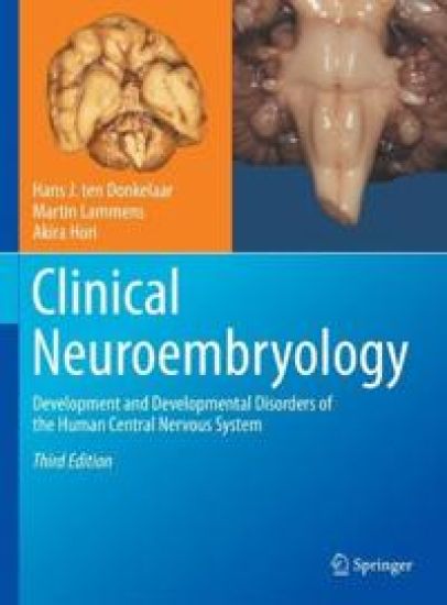
This book provides a comprehensive overview of the development of the human central nervous system (CNS) in the context of its many developmental disorders due to genetic, environmental and hypoxic/ischaemic causes.
Clinical Neuroembryology

Clinical Neuroembryology

Hans J. ten Donkelaar; Martin Lammens; Akira Hori

Springer International Publishing AG
2023
sidottu
This book provides a comprehensive overview of the development of the human central nervous system (CNS) in the context of its many developmental disorders due to genetic, environmental and hypoxic/ischaemic causes. The book contains three general, introductory chapters in which an overview of the development of the human brain and spinal cord, a summary of mechanisms of development as obtained in experimental studies in various invertebrates and vertebrates, and an overview of the causes of congenital malformations are presented. The developmental disorders of the human brain and spinal cord are presented in a regional, more or less segmental way, starting with neurulation and neural tube defects, and ending with developmental disorders of the cerebral cortex. These chapters are abundantly illustrated in colour with carefully chosen clinical case studies with imaging data, and when available, postmortem verification of the developmental disorders involved. In thethird edition, more emphasis has been given to the developmental ontology based on the prosomeric approach, and fetal development. Prenatal diagnosis by ultrasound, MRI and DTI, and classifications of developmental disorders have been updated. A number of new Clinical Cases have been included. Several new co-authors participate in various chapters. The book is intended for advanced medical students, and all those clinicians working with children and adults with developmental disorders of the CNS. Unique to the book is the integration of data from human embryology, experimental and molecular findings in mice in particular, imaging and developmental neuropathology.
Clinical Neuroanatomy

Clinical Neuroanatomy

Hans J. ten Donkelaar

Springer Nature Switzerland AG
2021
nidottu
Connections define the functions of neurons: information flows along connections, as well as growth factors and viruses, and even neuronal death can progress through connections. Accordingly, knowing how the various parts of the brain are interconnected to form functional systems is a prerequisite for properly understanding data from all fields in the neurosciences. Clinical Neuroanatomy: Brain Circuitry and Its Disorders bridges the gap between neuroanatomy and clinical neurology. It focuses on human and primate data in the context of brain circuitry disorders, which are so common in neurological practice. In addition, numerous clinical cases are presented to demonstrate how normal brain circuitry can be interrupted, and what the effects are. Following an introduction to the organization and vascularization of the human brain and the techniques used to study brain circuitry, the main neurofunctional systems are discussed, including the somatosensory, auditory,visual, motor, autonomic and limbic systems, the cerebral cortex and complex cerebral functions. In this 2nd edition, apart from a general updating, many new illustrations have been added and more emphasis is placed on modern techniques such as diffusion magnetic resonance imaging (dMRI) and network analysis. Moreover, a developmental ontology based on the prosomeric model is applied, resulting in a more modern subdivision of the brain. The new edition of Clinical Neuroanatomy is primarily intended for neurologists, neuroradiologists and neuropathologists, as well as residents in these fields, but will also appeal to (neuro)anatomists and all those whose work involves human brain mapping.
Clinical Neuroanatomy

Clinical Neuroanatomy

Hans J. ten Donkelaar

Springer Nature Switzerland AG
2020
sidottu
Connections define the functions of neurons: information flows along connections, as well as growth factors and viruses, and even neuronal death can progress through connections. Accordingly, knowing how the various parts of the brain are interconnected to form functional systems is a prerequisite for properly understanding data from all fields in the neurosciences. Clinical Neuroanatomy: Brain Circuitry and Its Disorders bridges the gap between neuroanatomy and clinical neurology. It focuses on human and primate data in the context of brain circuitry disorders, which are so common in neurological practice. In addition, numerous clinical cases are presented to demonstrate how normal brain circuitry can be interrupted, and what the effects are. Following an introduction to the organization and vascularization of the human brain and the techniques used to study brain circuitry, the main neurofunctional systems are discussed, including the somatosensory, auditory,visual, motor, autonomic and limbic systems, the cerebral cortex and complex cerebral functions. In this 2nd edition, apart from a general updating, many new illustrations have been added and more emphasis is placed on modern techniques such as diffusion magnetic resonance imaging (dMRI) and network analysis. Moreover, a developmental ontology based on the prosomeric model is applied, resulting in a more modern subdivision of the brain. The new edition of Clinical Neuroanatomy is primarily intended for neurologists, neuroradiologists and neuropathologists, as well as residents in these fields, but will also appeal to (neuro)anatomists and all those whose work involves human brain mapping.
An Illustrated Terminologia Neuroanatomica

An Illustrated Terminologia Neuroanatomica

Hans J. ten Donkelaar; David Kachlík; R. Shane Tubbs

Springer Nature Switzerland AG
2018
nidottu
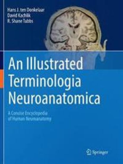
This book is unique in that it provides the reader with the most up-to-date terminology used to describe the human nervous system (central and peripheral) and the related sensory organs, i.e., the Terminologia Neuroanatomica (TNA), the official terminology of the IFAA (International Federation of Associations of Anatomists). The book provides a succinct but detailed review of the neuroanatomical structures of the human body and will greatly benefit not only various specialists such as (neuro)anatomists, neurologists and neuroscientists, but also students taking neuroanatomy and neuroscience courses. The book offers a high yield, combined presentation of neuroanatomical illustrations and text and provides the reader a ‘one-stop source’ for studying the intricacies of the human nervous system and its sensory organs. It includes an alphabetical list of official English terms and synonyms with the official Latin terms and synonyms from the TNA. With regard to the entries, the nameof the item in standardized English is provided, followed by synonyms and the official TNA Latin term, Latin synonyms and eponyms, a short description and in many cases one or more illustrations. To facilitate the use of illustrations, certain entries such as the gyri or sulci of the cerebral cortex are presented together with extensive cross-references. Terms that form part of a certain structure (such as the amygdaloid body, the thalamus and the hypothalamus) are listed under the respective structure. Segments and branches of arteries are discussed under the main artery, for example the A1–A5 segments under the anterior cerebral artery. Most nerves can be found following their origin from the brachial, cervical and lumbosacral plexuses. However, the major nerves of the limbs are discussed separately, as are the cranial nerves. Nuclei can be found by their English name or under Nuclei by their eponym.
An Illustrated Terminologia Neuroanatomica

An Illustrated Terminologia Neuroanatomica

Hans J. ten Donkelaar; David Kachlík; R. Shane Tubbs

Springer International Publishing AG
2018
sidottu
This book is unique in that it provides the reader with the most up-to-date terminology used to describe the human nervous system (central and peripheral) and the related sensory organs, i.e., the Terminologia Neuroanatomica (TNA), the official terminology of the IFAA (International Federation of Associations of Anatomists). The book provides a succinct but detailed review of the neuroanatomical structures of the human body and will greatly benefit not only various specialists such as (neuro)anatomists, neurologists and neuroscientists, but also students taking neuroanatomy and neuroscience courses. The book offers a high yield, combined presentation of neuroanatomical illustrations and text and provides the reader a ‘one-stop source’ for studying the intricacies of the human nervous system and its sensory organs. It includes an alphabetical list of official English terms and synonyms with the official Latin terms and synonyms from the TNA. With regard to the entries, the nameof the item in standardized English is provided, followed by synonyms and the official TNA Latin term, Latin synonyms and eponyms, a short description and in many cases one or more illustrations. To facilitate the use of illustrations, certain entries such as the gyri or sulci of the cerebral cortex are presented together with extensive cross-references. Terms that form part of a certain structure (such as the amygdaloid body, the thalamus and the hypothalamus) are listed under the respective structure. Segments and branches of arteries are discussed under the main artery, for example the A1–A5 segments under the anterior cerebral artery. Most nerves can be found following their origin from the brachial, cervical and lumbosacral plexuses. However, the major nerves of the limbs are discussed separately, as are the cranial nerves. Nuclei can be found by their English name or under Nuclei by their eponym.
Clinical Neuroembryology

Clinical Neuroembryology

Hans J. ten Donkelaar; Martin Lammens; Akira Hori

Springer-Verlag Berlin and Heidelberg GmbH Co. K
2016
nidottu
This book provides a comprehensive overview of the development of the human central nervous system (CNS) in the context of its many developmental disorders due to genetic, environmental, and hypoxic/ischemic causes. The introductory chapters give an overview of the development of the human brain and the spinal cord, the mechanisms of development as obtained in experimental studies of various invertebrates and vertebrates, and the causes of congenital malformations. In the main part, the developmental disorders of the human brain and the spinal cord are presented in a regional, more or less segmental way, starting with neurulation and neural tube defects, and ending with developmental disorders of the cerebral cortex. These are underlined by carefully chosen clinical case studies, including imaging data and, when available, postmortem verification of the developmental disorders involved. Numerous color photographs and illustrations complement the text. This second edition emphasizes the prenatal diagnosis by ultrasound, MRI, and DTI and implements new classifications of developmental disorders.
Clinical Neuroanatomy

Clinical Neuroanatomy

Hans J. ten Donkelaar

Springer-Verlag Berlin and Heidelberg GmbH Co. K
2016
nidottu
Connections define the functions of neurons: information flows along connections, as well as growth factors and viruses, and even neuronal death may progress through connections. Knowledge of how the various parts of the brain are interconnected to form functional systems is a prerequisite for the proper understanding of data from all fields in the neurosciences. Clinical Neuroanatomy: Brain Circuitry and Its Disorders bridges the gap between neuroanatomy and clinical neurology. It emphasizes human and primate data in the context of disorders of brain circuitry which are so common in neurological practice. In addition, numerous clinical cases demonstrate how normal brain circuitry may be interrupted and to what effect. Following an introduction into the organization and vascularisation of the human brain and the techniques to study brain circuitry, the main neurofunctional systems are discussed, including the somatosensory, auditory, visual, motor, autonomic andlimbic systems, the cerebral cortex and complex cerebral functions.
The Central Nervous System of Vertebrates

The Central Nervous System of Vertebrates

Rudolf Nieuwenhuys; Hans J. ten Donkelaar; Charles Nicholson

Springer-Verlag Berlin and Heidelberg GmbH Co. K
2014
nidottu
This comprehensive reference is clearly destined to become the definitive anatomical basis for all neuroscience research. The book provides a complete overview and comparison of the structural organization of all vertebrate groups, ranging from amphioxus and lamprey through fishes, amphibians and birds to mammals. The large specialised section of the work, devoted to the CNS of the various vertebrate groups, is preceded by introductory chapters on neurons, cell masses, fibre tracts, morphogenesis, methodology, and techniques. Although focusing on structure, the authors provide functional correlations throughout. This monumental work is, and will remain, unique; the only source of such brilliant illustrations at both the macroscopic and microscopic levels.
Clinical Neuroembryology

Clinical Neuroembryology

Hans J. ten Donkelaar; Martin Lammens; Akira Hori

Springer-Verlag Berlin and Heidelberg GmbH Co. K
2014
sidottu
This book provides a comprehensive overview of the development of the human central nervous system (CNS) in the context of its many developmental disorders due to genetic, environmental, and hypoxic/ischemic causes. The introductory chapters give an overview of the development of the human brain and the spinal cord, the mechanisms of development as obtained in experimental studies of various invertebrates and vertebrates, and the causes of congenital malformations. In the main part, the developmental disorders of the human brain and the spinal cord are presented in a regional, more or less segmental way, starting with neurulation and neural tube defects, and ending with developmental disorders of the cerebral cortex. These are underlined by carefully chosen clinical case studies, including imaging data and, when available, postmortem verification of the developmental disorders involved. Numerous color photographs and illustrations complement the text. This second edition emphasizes the prenatal diagnosis by ultrasound, MRI, and DTI and implements new classifications of developmental disorders.