Kirjojen hintavertailu – 12 903 725 kirjaa ja 27 kauppaa

Kirjailija

Hariqbal Singh

Kirjat ja teokset yhdessä paikassa: 20 kirjaa, julkaisuja vuosilta 2010–2024, suosituimpiin kuuluu Textbook of Radiology Physics. Vertaile teosten hintoja ja tarkista saatavuus suomalaisista kirjakaupoista.

20 kirjaa

Kirjojen julkaisuvuodet: 2010–2024.

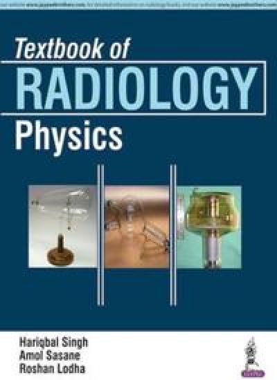
Textbook of Radiology Physics

Textbook of Radiology Physics

Hariqbal Singh; Amol Sasane; Roshan Lodha

Jaypee Brothers Medical Publishers
2016
nidottu
This book provides a concise overview of the field of radiology physics and its application in everyday practice. Beginning with an introduction to the fundamental concepts and the basics of radiation, the following sections review different techniques, from X-Ray production and ultrasound, to Doppler, mammography, computed tomography, and nuclear medicine procedures. Further topics include complex magnetic resonance concepts, radiation exposure monitoring, single-photon emission computed tomography, and positron emission tomography. Enhanced by radiological images and illustrations, each chapter explains the principles, function, application and limitations of the radiological technique in question. Key points Concise review of the field of radiology physicsCovers complete range of radiology techniques, from basic to more complexPrinciples, function, application and limitations of each technique explained in detailIncludes radiological images and illustrations to enhance learning
Love Radiology

Love Radiology

Hariqbal Singh; Mahesh Gautam; Robinson Shrestha

White Falcon Publishing
2022
sidottu
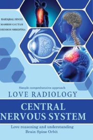
Love Radiology Central Nervous System" represents love and adoration towards learning Radiology. Once you stroll through this title, understand it, recognize it, you will adulate it, with an attempt to preserve a long-term lasting impact. This title is presented simple comprehensive approach to Brain Spine Orbit with ease of reading and understanding the subject with images, to acquire enough knowledge before appearing for the post graduate examination or reporting CNS cases more so on MR in order to arrive at most suitable conclusion.. This title is equally relevant for students appearing for the post graduate examination in clinical subjectsHope you will treasure reading and understanding this title. Hariqbal Singh Heads the Department of Radiology, Nobel Medical College. He has technical expertise, managing equipment, personnel and resources. An effective administrator. He has a large number of publications in countrywide and global medical journals. He has authored quite a few books on Imaging. He has been a post-graduate teacher, examiner and guide since 1989 with Maharashtra University of Health Sciences, Diplomate National Board, Savitribai Phule Pune University, Sumandeep University, BP Koirala Institute of Health Sciences (Nepal) and Kathmandu University. Mahesh Gautam is Assistant Professor in Department of Radiology, Nobel Medical College. He has several publications in medical journals. He is a profound and keen radiologist, enthusiastic and zealous observer. Along with Hariqbal Singh and Robinson Shrestha, he has authored Love Radiology series. Robinson Shrestha is Lecturer in Department of Radiology, Nobel Medical College. He is a disciplined, profound, reflective and ardent radiologist, fervent and passionate observer. Along with Hariqbal Singh and Mahesh Gautam, he has authored Love Radiology series.
Textbook of Radiology: Abdomen and Pelvis

Textbook of Radiology: Abdomen and Pelvis

Hariqbal Singh

Jaypee Brothers Medical Publishers
2017
nidottu
This book is a practical guide to the latest radiology techniques for the diagnosis of disorders of the abdomen and pelvis. Beginning with an overview of anatomy of the abdomen and pelvis, and a chapter explaining the features of children’s abdomens, the following sections cover the radiological aspects of different parts of the abdomen – gastrointestinal tract, hepatobiliary system, pancreas, renal system, adrenals, and retroperitoneum. The final chapters discuss imaging techniques for male reproductive organs, and for diagnosing obstetrics and gynaecological disorders. The text covers various imaging techniques – ultrasound, MRI, CT, X-Ray – and is highly illustrated with radiological images, diagrams and tables to enhance learning. Common exam questions are included to assist revision. Key Points Practical guide to latest radiological techniques for diagnosis of abdominal and pelvic disordersCovers various technologies and all parts of the abdomenIncludes imaging techniques for male reproductive organs and obstetrics and gynaecological disordersFeatures common exam questions to assist revision
Atlas of Human Anatomy on MRI

Atlas of Human Anatomy on MRI

Hariqbal Singh; Parvez Sheik

Jaypee Brothers Medical Publishers
2017
nidottu

Halvin toimitettuna 70,90 €

This book is a concise overview of MRI (magnetic resonance imaging) for brain, chest and abdominal disorders covering the very latest technologies and developments in the field. Beginning with an introduction to anatomy of these body systems, the following sections cover MR cholangiopancreatography, MRI of the female and male pelvis, and MR angiography. The atlas is enhanced by high quality MR images and tables with detailed descriptions to help clinicians understand complex anatomy. The comprehensive appendix provides a glossary of MRI terms and radiology measurement tables. Key Points Concise overview of MRI for brain, chest and abdomenFeatures sections on MR cholangiopancreatography, MRI of the pelvis, and MR angiographyComprehensive appendix provides glossary of terms and radiology measurement tablesIncludes high quality MR images and tables illustrating complex anatomy
Textbook of Radiology: Musculoskeletal Radiology

Textbook of Radiology: Musculoskeletal Radiology

Hariqbal Singh

Jaypee Brothers Medical Publishers
2016
nidottu
This book is a concise guide to musculoskeletal imaging for radiologists. Beginning with chapters on congenital skeletal anomalies and dysplasia, trauma, and metabolic and endocrine disorders, the following sections cover infections, arthritis, bone tumours, and disorders found in joints, soft tissues and breast. Each section covers both common and less common diseases and disorders and provides in depth discussion on the different imaging techniques including radiography, ultrasound, MRI, computed tomography, and nuclear magnetic resonance. The book is highly illustrated with nearly 200 radiological images and tables to enhance learning. Key Points Concise guide to musculoskeletal imagingCovers all modalities – radiography, MRI, CT, US and NMRDetailed discussion on diagnosis of both common and less common disorders and diseasesHighly illustrated with nearly 200 radiological images and tables
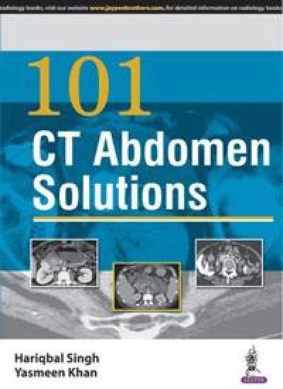
101 CT Abdomen Solutions

101 CT Abdomen Solutions

Hariqbal Singh; Yasmeen Khan

Jaypee Brothers Medical Publishers
2016
nidottu
101 CT Abdomen Solutions is a guide to interpreting computed tomography images of the abdominal anatomy. The book is divided into 23 sections, each covering every part of the abdomen from diaphragm and colon to prostate and uterus. This broad collection of CT images and high resolution axial CT images for all common pathologies assists interpretation and diagnosis in routine reporting practice. Enhanced by 162 high quality images and illustrations, 101 CT Abdomen Solutions is an effective reference guide for all radiologists. Key Points Comprehensive guide to interpreting CT images of the abdomen23 sections cover every part of the abdominal anatomy162 high quality images and illustrationsIncludes high resolution axial CT images for all common pathologies
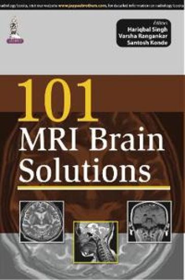
101 MRI Brain Solutions

101 MRI Brain Solutions

Hariqbal Singh; Varsha Angankar; Santosh Konde

Jaypee Brothers Medical Publishers
2015
nidottu
101 MRI Brain Solutions is a compilation of 101 cases related to congenital malformations and disorders affecting the brain. This comprehensive book details the use of MRI for diagnosis of infections including tuberculous meningitis, cerebral tuberculoma, multiple cerebral tuberculomas, viral encephalitis, Japanese encephalitis, neurocysticercosis, brain abscess and abscesses and ventriculitis. This book is divided into 12 chapters, with the first two discussing the basics of MRI and the anatomy of the brain. The following 9 chapters present a compilation of cases encountered in routine practice as well as more difficult cases. The last two chapters of this book are dedicated to the glossary of MRI Terms and MRI Acronyms. The use of MRI in the diagnosis of metachromatic leukodystrophy, Alexander disease, Canavan’s disease, adrenoleukodystrophy and multiple sclerosis, is highlighted with the help of clinical cases. 101 MRI Brain Solutions is an important resource for a broad audience, from post graduate students to practising radiologists and MR technical staff. Key Features Relevant to a wide audienceIncludes glossary of MRI terms and MRI acronymsChapter on MR anatomy of the brain166 full colour images and illustrations
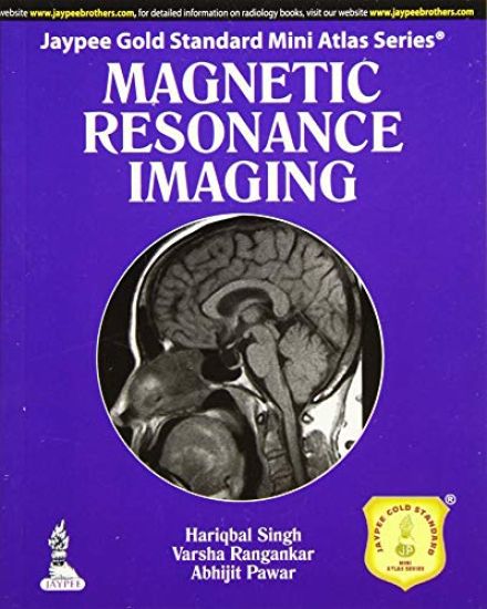
Jaypee Gold Standard Mini Atlas Series: Magnetic Resonance Imaging

Jaypee Gold Standard Mini Atlas Series: Magnetic Resonance Imaging

Hariqbal Singh; Varsha Rangankar; Abhijit Pawar

Jaypee Brothers Medical Publishers
2014
nidottu
Magnetic resonance imaging (MRI) is a type of scan that uses strong magnetic fields and radio waves to produce detailed images of the inside of the body. An MRI scanner is a large tube that contains powerful magnets. A patient lies inside the tube during the scan (NHS Choices). Part of the Jaypee Gold Standard Mini Atlas Series, this book is a comprehensive guide to MRI. Beginning with an introduction to the history and physics of MRI, each of the following sections discusses imaging and diagnosis of diseases in different areas of the body. MRI images for each condition are preceded by text summarising the disease, ultrasound and CT findings and important MRI findings to help clinicians interpret and make accurate diagnoses. The final sections list MR terms and acronyms. Key points Part of the Jaypee Gold Standard Mini Atlas SeriesComprehensive guide to Magnetic Resonance Imaging (MRI)Presents MRI images with descriptive text for numerous diseasesIncludes lists of key MR terms and acronyms
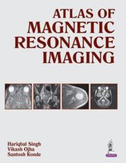
Atlas of Magnetic Resonance Imaging

Atlas of Magnetic Resonance Imaging

Hariqbal Singh; Vikash Ojha; Santosh Konde

Jaypee Brothers Medical Publishers
2014
sidottu
Magnetic resonance imaging (MRI) is a scan that uses strong magnetic fields and radio waves to produce detailed images of the inside of the body. This atlas is a comprehensive guide to MRI for radiology trainees and practising clinicians. Beginning with an introduction to the technique and the associated physics, each of the following chapters presents numerous high quality MRI images of different body systems including brain, orbit, spine, pelvis, and musculoskeletal system. Images are accompanied by detailed descriptions and each topic begins with a section on relevant anatomy. A self assessment chapter is included to test knowledge, and the final chapters include a glossary of MRI terms and MRI acronyms. Key points Comprehensive guide to MRI for trainees and radiologistsIn depth coverage of different body systemsTopics illustrated by high quality MRI images with descriptionsIncludes self assessment section
Jaypee Gold Standard Mini Atlas Series: Chest Radiology

Jaypee Gold Standard Mini Atlas Series: Chest Radiology

Hariqbal Singh

Jaypee Brothers Medical Publishers
2013
nidottu
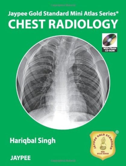
Chest radiology is the most commonly-used investigation method for pulmonary diseases. Accurate interpretation by radiologists is essential, in order to diagnose and manage disorders effectively. This atlas is a concise guide to chest radiology for residents and clinicians. Beginning with an introduction to anatomy, the book presents state of the art chest X-Ray, CT, MRI and PET scan images for numerous medical conditions. The book offers a clear understanding of how to recognise and interpret basic radiological signs, pathologies and patterns for differential diagnosis. A photo CD ROM is included to enhance the numerous well-illustrated X-Rays, CT and MRI scans in the atlas, making it an invaluable, hands-on reference for the evaluation of chest images. Key points Concise guide to chest radiology for residents and cliniciansPresents numerous chest X-Rays, CT, MRI and PET scan images for chest diseases and disordersOffers clear understanding of radiological signs, pathologies and patterns to assist differential diagnosisIncludes photo CD ROM
Atlas on X-Ray and Angiographic Anatomy

Atlas on X-Ray and Angiographic Anatomy

Hariqbal Singh; Parvez Sheik

Jaypee Brothers Medical Publishers
2013
nidottu
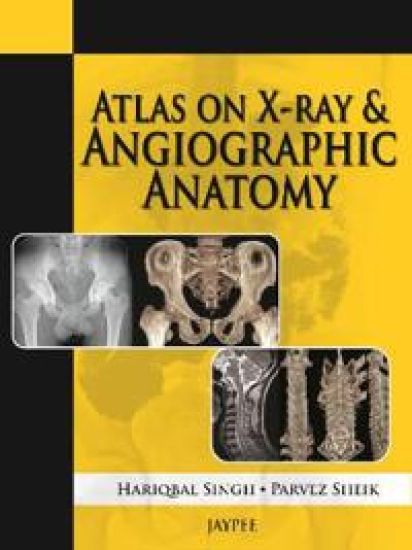
This atlas presents trainees with numerous X-ray and angiographic images to gain a thorough understanding of normal radiographic anatomy in order to make an accurate diagnosis of underlying pathology. Presented in an easy to read format, the book covers radiological procedures, ossification centres, X-ray production, digital subtraction angiography, and computed and digital radiography, in the different anatomical sections of the body. This practical guide includes nearly 240 clearly labelled images, illustrations and tables, with detailed descriptions, to assist learning. Key points Atlas of X-ray and angiographic images to help trainees understand normal radiographic anatomy and diagnose underlying pathologyEasy to read formatCovers different imaging techniques for all areas of the bodyIncludes nearly 240 images, illustrations and tables with detailed descriptions
101 Chest X-Ray Solutions

101 Chest X-Ray Solutions

Hariqbal Singh

Jaypee Brothers Medical Publishers
2013
nidottu
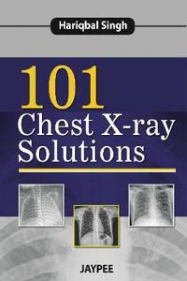
Chest X-ray is the most commonly requested film in any medical department so it is vital for clinicians to have a good understanding of the signs in order to make an accurate diagnosis. This book presents numerous postero-anterior plain chest X-ray images with detailed descriptions, exposing trainees and clinicians to both basic and more complex diagnoses. Beginning with an introduction to chest anatomy and how to read an X-ray, the following chapters present 101 cases by chest section. The final chapter includes 20 practice X-rays or spotters for self assessment. Key points 101 cases presenting chest X-ray images with detailed descriptionsCovers simple and complex diagnosesCases presented in logical format by section of the chestIncludes 20 practice X-rays for self assessment
Cross Sectional Imaging Made Easy

Cross Sectional Imaging Made Easy

Hariqbal Singh

Jaypee Brothers Medical Publishers
2011
nidottu
This book is intended to serve as comprehensive and easy-to-understand guide to cross sectional imaging. It presents principles of imaging modalities in a simple manner with cross sectional clinical illustrations and images to assist imaging departments. It is useful for medical students, junior doctors, residents, radiologists, colleagues in medicine, general practitioners, cross sectional imaging technical staff, medical colleges, institutional and departmental libraries, and for stand-alone cross sectional imaging establishments.
Practical Atlas of Computed Tomography

Practical Atlas of Computed Tomography

Hariqbal Singh; Sushil Kachewar

Jaypee Brothers Medical Publishers
2010
sidottu
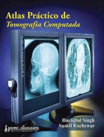
A systematic approach to Computed Tomographic imaging, this book contains normal anatomy, diverse pathologies and cross sectional anatomy to allow the specialist radiologist in practice or training to interpret and diagnose. The book is organised by body system and includes normal anatomy and a wide range of pathologies. Each clearly labelled image is accompanied by a reference image plane to allow ease of interpretation. Self assessment tools are also included.