Kirjojen hintavertailu – 12 903 733 kirjaa ja 27 kauppaa

Kirjailija

Joel A. Vilensky

Kirjat ja teokset yhdessä paikassa: 6 kirjaa, julkaisuja vuosilta 2010–2018, suosituimpiin kuuluu Netter's Concise Radiologic Anatomy Updated Edition. Vertaile teosten hintoja ja tarkista saatavuus suomalaisista kirjakaupoista.

6 kirjaa

Kirjojen julkaisuvuodet: 2010–2018.

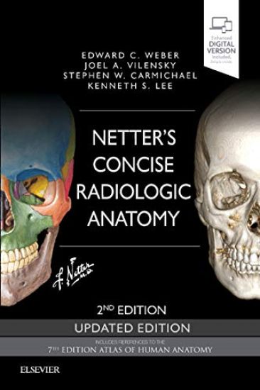
Netter's Concise Radiologic Anatomy Updated Edition

Netter's Concise Radiologic Anatomy Updated Edition

Edward C. Weber; Joel A. Vilensky; Stephen W. Carmichael

Elsevier - Health Sciences Division
2018
nidottu
Designed to make learning more interesting and clinically meaningful, Netter's Concise Radiologic Anatomy matches radiologic images?from MR and ultrasound to CT and advanced imaging reconstructions?to the exquisite artwork of master medical illustrator Frank H. Netter, MD. As a companion to the bestselling Netter's Atlas of Human Anatomy, this updated medical textbook begins with the anatomy and matches radiologic images to the anatomic images; the result is a concise, visual guide that shows how advanced diagnostic imaging is an amazing "dissection tool" for viewing human anatomy in the living patient! View direct, at-a-glance comparisons between idealized anatomic illustrations and real-life medicine with side-by-side radiology examples of normal anatomy and common variants with corresponding anatomy illustrations. Improve upon your knowledge with a brief background in basic radiology, including reconstructions and a list of common abbreviations for the images presented. Broaden your visual comprehension with the help of 30 brand-new ultrasound images. Access the complete contents online at Student Consult. NEW to this UPDATED EDITION: Cross-referenced to the 7th Edition Netter/Atlas of Human Anatomy
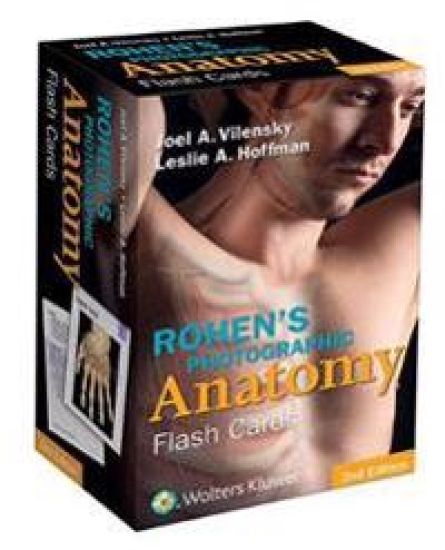
Rohen's Photographic Anatomy Flash Cards

Rohen's Photographic Anatomy Flash Cards

Joel A. Vilensky; Leslie A. Hoffman; Johannes W. Rohen; Chihiro Yokochi; Elke Lütjen-Drecoll

Lippincott Williams and Wilkins
2015
muu

Halvin toimitettuna 56,40 €

Derived from the leading dissection atlas Anatomy: A Photographic Atlas by Johannes W. Rohen, Chihiro Yokochi, and Elke Lütjen-Drecoll, these flash cards showcase full-color photographs of actual cadaver dissections. 220 cards, labeled for self-testing, serve as a practical tool to help students prepare for the dissection lab and the operating room. Now includes review questions and additional clinical anatomy.220 full-color photographs present exquisitely dissected cadavers. Ideal study aid to review recently dissected regions and to prepare for practical exams. Includes a key ring and hole-punched corners for easy organization and on-the-go portability.
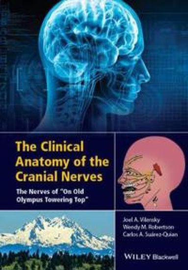
The Clinical Anatomy of the Cranial Nerves

The Clinical Anatomy of the Cranial Nerves

Joel A. Vilensky; Wendy Robertson; Carlo A. Suarez–Quian

John Wiley Sons Inc
2015
sidottu
The cranial nerves are an endlessly fascinating family of twelve nerves that have a dramatic impact on our daily lives. A dysfunction of the cranial nerves can cause loss of vision or double vision, loss of smell, poor balance, or loss of muscle function, and can also be an indicator of underlying neurological disorders. The Clinical Anatomy of the Cranial Nerves: The Nerves of "On Old Olympus Towering Top is an engaging and accessible book on the anatomy and clinical importance of these unique nerves. Thetext opens with a brief introduction of key neuroanatomical concepts that relate the clinical and anatomical sections that follow. Additionally, this book uniquely provides a detailed description of the bones of the head and face in order for the reader to understand the routes taken by the cranial nerves through the skull. Chapters then detail each nerve and its unique impact in relationship to our senses, motor function, and health. Vividly illustrated and supported by real-life clinical cases, the book will appeal to anyone wishing to gain a better understanding of the cranial nerves. Merging anatomical and clinical information with intriguing clinical cases, The Clinical Anatomy of the Cranial Nerves: The Nerves of "On Old Olympus Towering Top introduces readers to the anatomy and diverse function of this intriguing family of nerves.
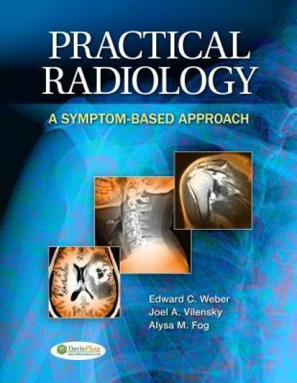
Practical Radiology

Practical Radiology

Edward C. Weber; Joel A. Vilensky; Alysa Fog

F.A. Davis Company
2013
nidottu
Rely on this practical guide to the role of medical imaging in the diagnosis and treatment of common diseases and disorders. Follow its symptoms-based approach to learn when medical imaging is appropriate, what the ideal study may be for a specific clinical problem, how to interpret an official report on a radiologic study, what the possible appropriate next steps are, and how radiologic results may (or may not) alter clinical management of your patient. Case studies in each chapter present typical patients with accompanying radiologic reports. Actual images of superb clarity show you the potential of contemporary techniques. Master the basics of medical imaging in patient care, the physical principles underlying imaging modalities- including conventional radiology, CT, MRI, ultrasound, and nuclear medicine scans- as well as common pitfalls.
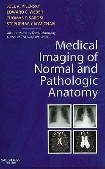
Medical Imaging of Normal and Pathologic Anatomy

Medical Imaging of Normal and Pathologic Anatomy

Joel A. Vilensky; Edward C. Weber; Thomas Sarosi; Stephen W. Carmichael

W B Saunders Co Ltd
2010
nidottu
Written for the modern medical student and designed to accompany any current gross anatomy textbook, this brand-new pictorial handbook presented by Drs. Vilensky, Weber, Carmichael, and Sarosi lets you quickly identify pathologic correlates of gross anatomy. Abundant side-by-side high-quality radiography, MR, CT, and ultrasound images of normal and pathologic conditions help you quickly develop the skills you need to differentiate between what's normal and what's not. Discussions on the choice of imaging modality for various pathologies will help you select the right imaging procedure in many clinical situations, making this a handy resource in the clinical environment. But best of all, this visual approach to pathologic correlates will help you ace your courses, the USMLE and NBME final exams. . Features side-by-side radiography, MR, CT, and ultrasound images that illustrate normal and abnormal anatomy, helping you quickly identify conditions while improving your diagnostic skills. . Covers clinical conditions found in the main core of textbooks and radiologically depicts the clinical correlates that you're exposed to daily, making it the ideal companion resource for any medical gross anatomy course. . Uses concise, brief text that explains the condition, thus allowing the radiologic images to guide you to the differentiating factors. . Incorporates discussions of imaging modality choices for a range of pathologies to help you understand how to select imaging procedures for various clinical situations in the clinical environment. . Offers the visual guidance you need to study for and pass your exams.