

Kirjojen hintavertailu – 12 903 725 kirjaa ja 27 kauppaa

Kirjailija

Larry R. Cochard

Kirjat ja teokset yhdessä paikassa: 3 kirjaa, julkaisuja vuosilta 2011–2026, suosituimpiin kuuluu Netter's Embryology Coloring Book. Vertaile teosten hintoja ja tarkista saatavuus suomalaisista kirjakaupoista.

3 kirjaa

Kirjojen julkaisuvuodet: 2011–2026.

Netter's Embryology Coloring Book

Netter's Embryology Coloring Book

Larry R. Cochard; Angelique N. Dueñas

Elsevier Health Sciences
2026
nidottu
An all-new addition to the Netter Coloring Books family, Netter’s Embryology Coloring Book makes learning embryology easier and more enjoyable through active and engaging review! The study of embryology is particularly suited for coloring exercises because the actions of coloring and tracing reinforce spatial understanding and changes following the development of structures over time. With artwork based on the highly regarded and familiar Netter collection, you know you’re benefiting from reliable, up-to-date anatomical information. This new Netter coloring book also contains review questions and clinical notes, making it an excellent, high-yield embryology resource for foundational learning as well as review. Netter's Embryology Coloring Book is a perfect companion to Netter’s Anatomy Coloring Book as well as Netter’s Atlas of Human Embryology, Netter’s Physiology Coloring Book, Netter’s Neuroscience Coloring Book, Netter’s Anatomy Flash Cards, and Netter’s Physiology Flash Cards. Emphasizes the continuity between the primordia (precursors) in the early embryo and their derivatives in the fetus and adult Uses standard color coding for the three early embryo (gastrula) germ layers of cells: ectoderm blue, endoderm yellow, and mesoderm red reflect common conventions used in life sciences education Features Clinical Notes throughout that emphasize important aspects of development in a clinical context Includes review questions at the end of each section for convenient self-assessment and review Presents each topic in two-page spreads?with outlines of Netter anatomical illustrations for coloring on one side; and high-yield information and coloring guide on the other Features illustrations small enough for quick coloring, but large enough to provide important details Offers tips for coloring key structures that emphasize how a coloring exercise can reinforce learning Provides online access where you can view completed coloring pages for reference An eBook version is included with purchase. The eBook allows you to access all of the text and figures, with the ability to search, make notes and highlights, and have content read aloud

Netter's Atlas of Human Embryology

Netter's Atlas of Human Embryology

Larry R. Cochard; Angelique N. Dueñas

Elsevier Health Sciences
2024
nidottu

Halvin toimitettuna 71,90 €

Illustrated by the world-renowned Frank H. Netter, MD and artists working in his tradition, Netter's Atlas of Human Embryology, Second Edition, provides a rich pictorial overview of human prenatal development. For each stage of development and for each body system, you'll find a brief description of the developmental plan, with key concepts and terminology, followed by discussions of histological principles, the classification of congenital anomalies, and basic cellular, molecular, and genetic concepts. An emphasis on morphological patterns in the embryo and fetus makes it easy to understand the structure and function of the adult body and the embryonic basis of natural variation as well as birth anomalies. Uses vibrant, high-quality illustrations and concise text to teach key points quickly, effectively, and memorably Includes new Inclusionary Consideration Points that detail key information such as differences between sex and gender-particularly for the embryology of sex determination; and perspectives on disability and disability advocacy-including person-first vs. identity-first language; plus new illustrations with more diversity and better representation of skin tones, sex, and body habitus Discusses topics new to this edition, including Conjoined Twins, Early Detection of Pregnancy, Exstrophy of the Bladder, Genetic Sex Variations, Congenital Limb Anomalies, Holoprosencephaly, and more Provides expanded coverage of clinical correlations and congenital anomalies and new Clinical Notes boxes throughout Contains summary tables and terminology sections at the end of each chapter, plus an appendix with all major congenital anomalies and their embryonic basis, making it easy to review course material and prepare for the USMLE An eBook version is included with purchase. The eBook allows you to access all of the text, figures and references, with the ability to search, make notes and highlights, and have content read aloud Evolve Instructor site with an image collection is available to instructors through their Elsevier sales rep or via request at https://evolve.elsevier.com.
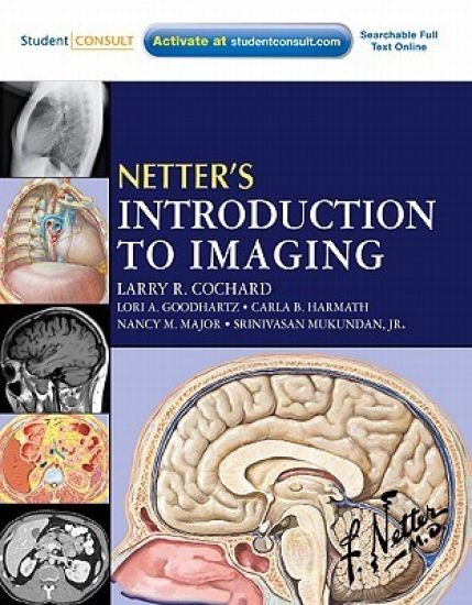
Netter's Introduction to Imaging

Netter's Introduction to Imaging

Larry R. Cochard; Lori A Goodhartz; Carla Harmath; Nancy M. Major; Srinivasan Mukundan

W B Saunders Co Ltd
2011
nidottu
Netter's Introduction to Imaging, by Larry R. Cochard, PhD, Lori A Goodhartz, MD Carla B, Harmath, MD, Nancy M. Major MD, and Srinivasan Mukundan, JR, MD, makes interpreting normal and abnormal X-ray, CT, and MR images easy by correlating them with crystal-clear Netter illustrations. You'll learn to recognize anatomical relationships in images and apply them to a variety of examples of pathology throughout the body, including the imaging of masses, air, or blood in organs and spaces...fractures, thickening, constriction, and compression...and more. You can also access the book from any computer at studentconsult.com. It's an ideal introduction to diagnostic imaging! Visualize anatomical structures and relationships with perfect clarity with the aid of vivid, colorful Netter artwork. The coloring, texture, and idealized emphasis help you interpret relationships between structures and compartments as seen in cross section and in X-rays, CT, and MRI. Develop your ability to better identify pathologies by viewing normal healthy anatomical images and abnormal images. Comparative images reinforce your basic understanding of what normal tissues and anatomy look like and serve as a guide in recognizing disease patterns and processes: atypically large or small organs and compartments, masses, air, or blood in organs and spaces, fractures, thickening, constriction, compression, and more. Understand the principles that underlie X-ray, CT, MR, ultrasound, and nuclear medicine imaging, the use of contrast and angiography. Understand how radiologists apply systematic search strategies in imaging studies of each region of the body. Access the complete text and images online at studentconsult.com