Kirjojen hintavertailu – 12 881 293 kirjaa ja 26 kauppaa

Kirjailija

Leslie H. Sobin

Kirjat ja teokset yhdessä paikassa: 5 kirjaa, julkaisuja vuosilta 1989–1996, suosituimpiin kuuluu Histological Typing of Intestinal Tumours. Vertaile teosten hintoja ja tarkista saatavuus suomalaisista kirjakaupoista.

5 kirjaa

Kirjojen julkaisuvuodet: 1989–1996.

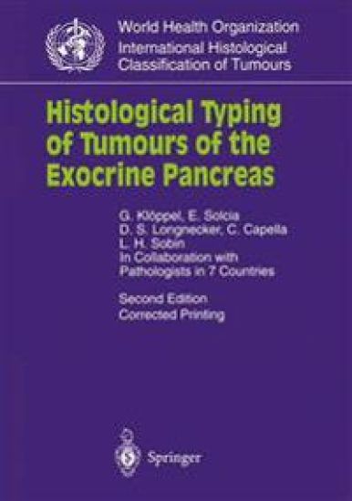
Histological Typing of Tumours of the Exocrine Pancreas

Histological Typing of Tumours of the Exocrine Pancreas

G. Kloppel; E. Solcia; D.S. Longnecker; C. Capella; Leslie H. Sobin

Springer-Verlag Berlin and Heidelberg GmbH Co. K
1996
nidottu
This is a histological classification of tumours and tumour-like lesions of the exocrine pancreas which also includes those tu- mours showing a mixture of exocrine and endocrine elements. The classification is based principally on standard microscopic observations, but whenever indicated it incorporates diagnosti- cally valuable immunohistological findings. In addition, the most important immunohistological findings which are helpful in cat- egorizing pancreatic tumours are summarised in Table 1. The major guideline of this classification scheme is the group- ing of the pancreatic exocrine tumours according to their biologi- cal behaviour. Thus, the neoplasms are broadly divided into benign (adenoma) and malignant tumours (carcinoma). How- ever, in recent years we have learned that this division is not a sharp but rather a gradual transition. We therefore added a third group which we call "tumours of uncertain malignant potential" representing a borderline category analogous to that recognized for some ovarian tumours. This group includes mucinous cystic tumour, intraductal papillary-mucinous tumour and solid- pseudopapillary tumour. These neoplasms are defined by the grade of dysplasia and/or potential to become malignant. Mucinous cystic tumours of uncertain malignant potential, for instance, exhibit moderate epithelial dysplasia, but do not show severe dysplasia/carcinoma in situ changes, nor carcinomatous invasion of the cyst wall or the adjacent pancreatic tissue. Solid- pseudopapillary tumour has a benign looking histological ap- pearance, but metastases may occur. Biologically, all these neoplasms are primarily slow-growing lesions and have an excel- lent prognosis when adequately treated by complete resection.
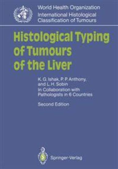
Histological Typing of Tumours of the Liver

Histological Typing of Tumours of the Liver

Kamal G. Ishak; Peter P. Anthony; Leslie H. Sobin

Springer-Verlag Berlin and Heidelberg GmbH Co. K
1994
nidottu
This is a classification of tumours and tumour-like lesions of the liver. It is based primarily on the microscopic characteristics of the tumours, and is therefore concerned with morphologically identi- fiable cell types and histological patterns. The haematoxylin- and eosin-stained section remains the mainstay of morphological diag- nosis, but special histochemical stains are often helpful and have been referred to in the explanatory notes. Readers interested in specific special stains mentioned in the text should consult Labora- tory Methods in Histotechnology of the Armed Forces Institute of *Pathology, Washington, D. c., USA. 1 The results of immunohisto- chemical methods for identifying various tumour "markers" have also been noted whenever indicated. The present classification incorporates all the previously classi- fied tumours, but also includes several new lesions, viz. biliary papil- lomatosis, the fibrolamellar variant of hepatocellular carcinoma and epithelioid haemangioendothelioma. Several subtypes of hepato- blastoma are mentioned. A serous type of bile duct cystadenoma is described. The section on tumour-like lesions has been expanded to include focal fatty change and inflammatory pseudotumour. The section on adenomatous hyperplasia, including macroregenerative nodules, has been amplified. The number of photomicrographs has been increased from the original 56 to 150. Unlike the first edition, the photomicrographs in the second edition are mostly black and white. All are new and were taken of representative cases on file at the Armed Forces Institute of Pathology.
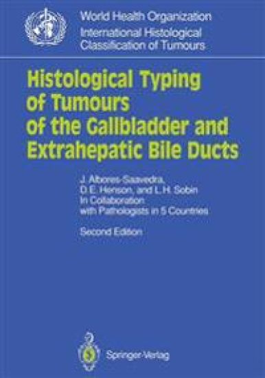
Histological Typing of Tumours of the Gallbladder and Extrahepatic Bile Ducts

Histological Typing of Tumours of the Gallbladder and Extrahepatic Bile Ducts

Jorge Albores-Saavedra; D.E. Henson; Leslie H. Sobin

Springer-Verlag Berlin and Heidelberg GmbH Co. K
1991
nidottu
This is a classification of tumours and tumour-like lesions of the gall- bladder and extrahepatic bile ducts, including the ampulla of Vater. Although most of the lesions are found in all three sites, variations in frequency of the histological types occur and will be noted. The incidence of carcinoma of the gallbladder varies in different parts of the world. Variation is also found in different ethnic groups within the same country. In the United States, for example, carcino- ma of the gallbladder is more common in American Indians than in Caucasians or in Blacks; the rate among female American Indians is 21 per 100000 compared with 1.4 per 100000 among Caucasian fe- males. In Latin American countries, the highest rates are found in Chile, Mexico and Bolivia. In other countries, such as Japan, the inci- dence rates are intermediate between those of American Indians and those of Caucasians. Despite certain features in common, carcinomas of the gallblad- der and carcinomas of the extrahepatic bile ducts show a number of differences. Gallbladder carcinomas are usually associated with cholelithiasis and have a strong female predominance. In contrast, extrahepatic bile duct carcinomas are seen less often, occur in both sexes with equal frequency, are usually not associated with choledo- cholithiasis, produce early biliary obstruction, and are better differen- tiated histologically as a group. Moreover, they are seen in patients with primary sclerosing cholangitis and ulcerative colitis.
Histological Typing of Oesophageal and Gastric Tumours

Histological Typing of Oesophageal and Gastric Tumours

Hidenobu Watanabe; Jeremy R. Jass; Leslie H. Sobin

Springer-Verlag Berlin and Heidelberg GmbH Co. K
1990
nidottu
This classification is based primarily on the microscopic characteris- tics of tumours. It is therefore concemed with the identification of cell types and histological pattems as seen by conventional light mi- croscopy. In general, time-honoured terms have been retained. Syn- onyms are listed only if they have been widely used, or if they are considered to be important for understanding the disease process. In such cases the preferred term is given first, followed by the synonym in parentheses. The individuality of the tumour manifests itself principally in its histological appearance and the extent of spread at the time of diag- nosis. This volume is concemed only with the histological classifica- tion of tumours. Anatomieal extent or staging is covered in the TNM 1 Classification. The histological classification of a tumour depends on two main parameters, typing and grading, and a number of additional parame- ters which may apply to selected examples.
Histological Typing of Intestinal Tumours

Histological Typing of Intestinal Tumours

Jeremy R. Jass; Leslie H. Sobin

Springer-Verlag Berlin and Heidelberg GmbH Co. K
1989
nidottu
This classification is based primarily on the microscopic characteris­ tics of tumours. It is therefore concerned with the identification of cell types and histological patterns as seen by conventional light micro­ scopy. In general, time-honoured terms have been retained. Syn­ onyms are listed only if they have been widely used or if they are con­ sidered to be important for understanding the disease process. In such cases, the preferred term is given first, followed by the synonym in parentheses. The individuality of a tumour manifests itself principally in its histological appearance and the extent of spread at the time of diag­ nosis. This volume is concerned only with the histological classific­ ation of tumours. Anatomical extent or staging is covered in the TNM Classification.! The histological classification of a tumour de­ pends on two main parameters, typing and grading, and a number of additional parameters which may apply to selected examples. Histological Typing This divides tumours of a given organ into different types according to their direction of differentiation. Although this may frequently in­ dicate the underlying histogenesis of the tumour, it may be difficult or impossible to identify the cell of origin. Note is taken of the structure and function of cell types, as well as the overall growth pattern of the tumour, with the aim of matching these features to those of a normal tissue found in the same organ.