Kirjojen hintavertailu – 12 903 733 kirjaa ja 27 kauppaa
Kirjailija
Marios Loukas
Kirjat ja teokset yhdessä paikassa: 11 kirjaa, julkaisuja vuosilta 2013–2024, suosituimpiin kuuluu GRAY'S Klinischer Fotoatlas Anatomie. Vertaile teosten hintoja ja tarkista saatavuus suomalaisista kirjakaupoista.
**Selected for 2025 Doody’s Core Titles® in Anatomy/Embryology** The perfect hands-on reference, Gray's Clinical Photographic Dissector of the Human Body, 3rd Edition, is a practical resource in the anatomy lab, on surgical rotations, during clerkship and residency and beyond! This fully revised third edition uses a unique, step-by-step presentation of full-color cadaveric photographs to orient you more quickly in the anatomy lab, and points out the clinical relevance of each structure and every dissection. Each photograph depicts clearly labeled anatomical structures, including muscles, bones, nerves, blood vessels, and organs-making this one-of-a-kind resource ideal for preparing for laboratory sessions and as a useful reference during dissections. Contains nearly 1,100 full-color photographs for comparison to the cadavers you study, helping you become more proficient and confident in your understanding of the intricacies of the human body Guides you through each dissection step-by-step, using a unique, real-world photographic presentation Includes complementary high-quality schematic drawings throughout to help orientate you and aid understanding Contains superb corresponding Gray’s illustrations to add clarity to key anatomical structures Helps you easily relate anatomical structures to clinical conditions and procedures Features new explanatory videos of human cadaveric dissection for each chapter Depicts the pertinent anatomy for more than 30 common clinical procedures such as prosthetic hip replacements, intravenous catheters, lumbar puncture, and knee joint aspiration, including where to make the relevant incisions Reflects the same level of accuracy and thoroughness that has made the Gray’s ‘family’ of products the most trusted learning resources in anatomy Prepared by an expert author team-highly experienced educators and leading authorities in clinical anatomy An eBook version is included with purchase. The eBook allows you to access all of the text and figures, with the ability to search, customize your content, make notes and highlights, and have content read aloud The Evolve Instructor site with downloadable images is available to instructors through their Elsevier sales rep or via request at https://evolve.elsevier.com
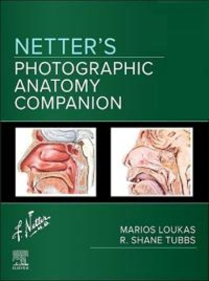
Connect what you see in prosections and dissections in the anatomy lab with the detailed medical illustrations of Frank H. Netter, MD. Netter's Photographic Anatomy Companion features 80 carefully selected plates from the well-known Netter Atlas of Human Anatomy, each paired with photographs of real cadaver prosections in the same position, plane, and view. This unique reference and study tool provides a visual connection between the idealized anatomical concepts in Netter’s famous illustrations with the photos of body donors seen in the anatomy lab, making it an invaluable resource for learning human anatomy, as a dissection reference, for practical exam review, and for prepping for surgical rotations. Provides a unique, visual connection between real-world cadaveric presentation in the anatomy lab with Netter Atlas of Human Anatomy, the most widely used anatomy atlas for medical programs. Pairs dissection photographs side by side with classic Netter illustrations in the same view and orientation, helping solve the common problem of dissected and prosected specimens of body donors that do not closely resemble the idealized images in an illustrated atlas. Makes it easier to locate structures in a body donor and connect that structure with the detailed, high-quality illustration of a didactic Netter plate, enabling you to more quickly discover, learn, and understand anatomical structures and relationships. An ideal reference tool for the prosection or dissection lab, anatomy instructors, and medical and allied health students, as well as an excellent study aid outside the lab for practicals, exams, and surgical rotations. An eBook version is included with purchase. The eBook allows you to access all of the text, figures, and references, with the ability to search, make notes and highlights, and have content read aloud.
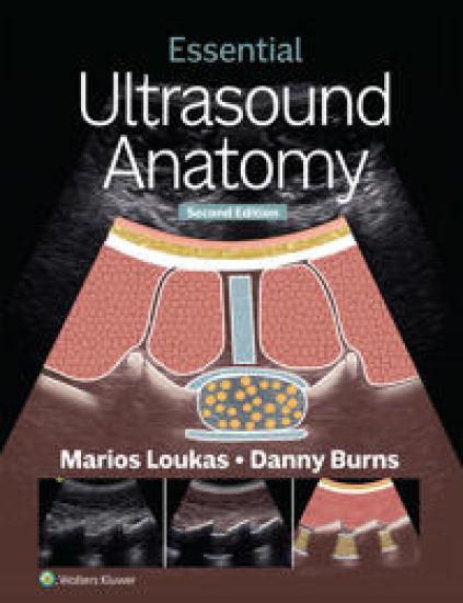
Ultrasound Anatomy, 2nd Edition, offers practical, comprehensive coverage of the ultrasound images and important structures that are most frequently encountered in daily practice and during point-of-care ultrasound (POCUS). This approachable text integrates concise text, detailed ultrasound images with corresponding cadaver photographs, full-color anatomical and technique illustrations, and videos to provide a solid foundation in regional ultrasound anatomy. The enhanced 2nd Edition features reorganized content for improved learning, with chapter Q&As now conveniently placed at the end of each section. More than 30 new figures, additional pathologies, and new content on central venous catheterization and joint aspiration make this an indispensable resource for today's ultrasound practitioners. NEW! Content on central venous catheterization and joint aspiration broaden students’ mastery of clinical sonography practices. UPDATED! Photos, illustrations, and ultrasound images clarify patient positioning, probe positioning, and surface anatomy in vibrant detail. Clinical Applications provide a clinical context for the types of structures and cases students will encounter on rotations. Focused Assessment with Sonography for Trauma (FAST) exam chapter familiarizes students with the protocols used by surgeons, emergency physicians, and some paramedics to screen for certain conditions after trauma. Ultrasound videos and USMLE-style review questions available online reinforce critical content and strengthen test-taking confidence.
With the most extensive, comprehensive collection of anatomy multiple-choice questions in strict, current USMLE format, Gray's Anatomy Review, 3rd Edition, is an easy-to-use study tool that helps you relate anatomy to clinical practice and pass your exams. Whether used as a companion to Gray's Anatomy for Students or as a stand-alone resource, this medical textbook is your indispensable review book for both in-course examinations and the USMLE Step 1. Includes more than 1,400 high-yield questions, mirroring the USMLE Step 1 and complete with answers and rationales, that challenge your grasp of anatomical knowledge and the anatomical basis of disease. Features a new neuroanatomy chapter containing approximately 100 all-new questions that cover key concepts and relate them to clinical practice. Groups questions more logically within chapters for more effective study, first within topic areas and then from easy to more difficult. Provides specific, updated page references to current editions of Gray's Anatomy for Students, plus key answers and explanations to Gray's Basic Anatomy and Gray's Atlas of Anatomy for additional review. Helps you visualize key concepts with updated radiographic and ultrasound images and extensive use of photographs. Enhanced eBook version included with purchase. Your enhanced eBook allows you to access interactive quizzes, plus all of the text, figures, and references from the book on a variety of devices.
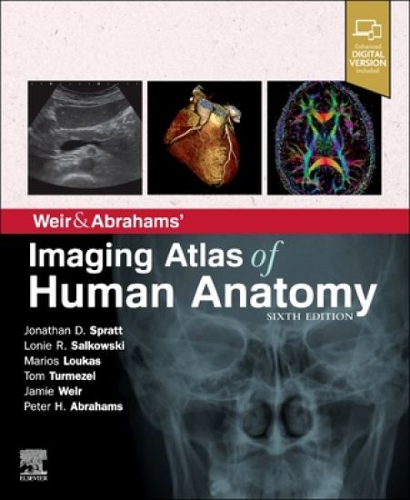
BMA Book Awards - Winner of Basic and Clinical Sciences category! The perfect up-to-date imaging guide for a complete and 3-dimensional understanding of applied human anatomy Imaging is ever more integral to anatomy education and throughout modern medicine. Building on the success of previous editions, this fully revised sixth edition provides a superb foundation for understanding applied human anatomy, offering a complete view of the structures and relationships within the whole body, using the very latest imaging techniques. All relevant imaging modalities are included, from plain radiographs to more advanced imaging of ultrasound, CT, MRI, functional imaging and angiography. Coverage is further enhanced by a carefully selected range of BONUS electronic content, including clinical photos and cases, ultrasound videos, labelled radiograph 'slidelines', cross-sectional imaging stacks and test-yourself materials. Uniquely, key syllabus image sets are now highlighted throughout to aid efficient study, as well as the most common, clinically important anatomical variants that you should be aware of. This superb package is ideally suited to the needs of medical students, as well as radiologists, radiographers and surgeons in training. It will also prove invaluable to the range of other students and professionals who require a clear, accurate, view of anatomy in current practice. Fully revised legends and labels and new high-quality images-featuring the latest imaging techniques and modalities as seen in clinical practice Covers the full variety of relevant modern imaging-including cross-sectional views in CT and MRI, angiography, ultrasound, fetal anatomy, plain film anatomy, nuclear medicine imaging and more - with better resolution to ensure the clearest anatomical views Core syllabus image sets now highlighted throughout-to help you focus on the most essential areas to excel on your course and in examinations Unique summaries of the most common, clinically important anatomical variants for each body region-reflects the fact that around 20% of human bodies have at least one clinically significant variant New orientation drawings-to help you understand the different views and the 3D anatomy of 2D images, as well as the conventions between cross-sectional modalities Ideal as a stand-alone resource or in conjunction with Abrahams' and McMinn's Clinical Atlas of Human Anatomy-where new links help put imaging in the context of the dissection room Now a more complete learning package than ever before, with superb BONUS electronic enhancements embedded within the accompanying eBook, including: Labelled image 'stacks'-that allow you to review cross-sectional imaging as if using an imaging workstation Labelled image 'slidelines'-showing features in a full range of body radiographs to enhance understanding of anatomy in this essential modality Self-test image 'slideshows' with multi-tier labelling-to aid learning and cater for beginner to more advanced experience levels Labelled ultrasound videos-bring images to life, reflecting this increasingly clinically practiced technique Questions and answers accompany each chapter-to test your understanding and aid exam preparation 34 pathology tutorials-based around nine key concepts and illustrated with hundreds of additional pathology images, to further develop your memory of anatomical structures and lead you through the essential relationships between normal and abnormal anatomy High-yield USMLE topics-clinical photos and cases for key topics, linked and highlighted in chapters
A unique biographical review of the global contributors to field of anatomy Knowledge of human anatomy has not always been an essential component of medical education and practice. Most European medical schools did not emphasize anatomy in their curricula until the post-Renaissance era; current knowledge was largely produced between the 16th and 20th centuries. Although not all cultures throughout history have viewed anatomy as fundamental to medicine, most have formed ideas about the internal and external mechanisms of the body?influences on the field of anatomy that are often overlooked by scholars and practitioners of Western medicine. History of Anatomy: An International Perspective explores the global and ancient origins of our modern-day understanding of anatomy, presenting detailed biographies of anatomists from varied cultural and historical settings. Chapters organized by geographic region, including Africa, the Middle East, and Europe, review the lives of those that helped shape our current understanding of the human form. Examining both celebrated and lesser-known figures, this comprehensive work examines their contributions to the discipline and helps readers develop a global perspective on a cornerstone of modern medicine and surgery. Offers a comprehensive and multidisciplinary examination of the history of anatomyTraces the emergence of modern knowledge of anatomy from ancient roots to the modern eraFills a gap in current literature on global perspectives on the history of anatomyWritten by an internationally recognized team of practicing physicians and scholars History of Anatomy: An International Perspective is an engaging and insightful historical review written for anatomists, anthropologists, physicians, surgeons, medical personnel, medical students, health related professionals, historians, and anyone interested in the history of anatomy, surgery, and medicine.
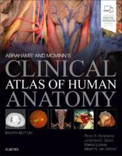
Abrahams' and McMinn's Clinical Atlas of Human Anatomy, 8th Edition delivers the straightforward visual guidance you need to perform confidently in all examinations and understand spatial relationships required during your medical training, while also acquiring the practical anatomical knowledge needed for your future clinical career. Respected authority Prof. Peter Abrahams and his team of leading international anatomists and radiologists link a vast collection of clinical images to help you master all the essential correlations between the basic science of anatomy and its clinical practice. See what to look for and how to proceed thanks to an unsurpassed collection of labelled dissection photographs, supported by clear, explanatory diagrams and modern imaging Correlate anatomy to clinical practice with a wealth of MR, CT, DSA, radiographic, endoscopic, and operative images that demonstrate how structures are viewed today in the clinical setting Thoroughly revised and updated throughout, including: brand new dissections, to further improve clarity and consistency throughout the book in every region all new colour overlays added to selected dissections making it even easier to identify key nerves, arteries, veins and especially lymphatics fully revised neuroanatomy content reflects the latest understanding of functional neuroanatomy as seen with modern 3D and functional imaging updated and coloured and a unique lymphatics section Includes access to the complete, enhanced eBook - unlocking a huge, carefully-selected collection of BONUS CLINICAL material, integrated throughout on almost every page to enhance your study and help make those essential clinical / anatomical connections. This sets Abrahams' and McMinn's apart from any other atlases of human anatomy! 200+ 3D scans,- allowing you to view the body in a more dynamic way to aid your understanding of dynamic anatomy. Master the 500 clinical conditions every physician should know by reviewing the associated clinical topics - featuring over 2000 additional clinical photos, radiological images, and case presentations not found in the printed book. Learn from the culmination of over 45 years international clinical experience of Prof. Abrahams and over 100 of his colleagues worldwide who have contributed to this unique collection of clinical anatomy images.
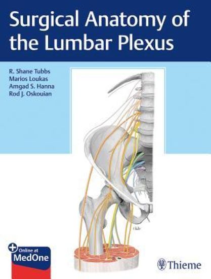
Dedicated guide to the lumbar plexus provides invaluable anatomical and surgical insights Thorough knowledge of the lumbar plexus and its branches is crucial to achieving positive patient outcomes, especially with newer surgical approaches. Many of the nerve branches are formed within the psoas major muscle and careful dissection is necessary to free them during surgery to prevent damage. Moreover, the iliac vessels are medial to some of the larger branches of the plexus, such as the femoral and obturator nerves. In the retroperitoneal space, the kidney and ureter are nearby. In addition, due to the overlying peritoneal cavity and its contents, accessing the lumbar plexus presents considerable challenges. Surgical Anatomy of the Lumbar Plexus is the only book on the market devoted to the lumbar plexus and its branches, focusing on anatomy and clinical applications, pathology, surgery, and imaging. Internationally known authors R. Shane Tubbs, Marios Loukas, Amgad Hanna, Rod Oskouian and a cadre of esteemed specialists provide unique insights, clinical pearls, knowledge based on thousands of spine surgeries, and a well-rounded multidisciplinary perspective. Key HighlightsNine separate chapters dedicated to impacted nerves: subcostal, iliohypogastric, ilioinguinal, lateral femoral cutaneous, genitofemoral, femoral, obturator, furcal, and accessory obturatorHistory and overview of dermatomal anatomy of branches derived from the lumbar plexusAnesthetic blockade of the femoral, lateral femoral cutaneous, iliohypogastric, ilioinguinal, and genitofemoral nervesTips for preventing complications and achieving positive outcomes for lateral transpsoas approaches to the lumbar spine and other proceduresMore than 100 novel illustrations provide clarity and understanding of a complex anatomical area as it relates to surgery involving the lumbar plexus and branches This o
Written with the student in mind, Netter's Introduction to Clinical Procedures, by Drs. Marios Loukas, R. Shane Tubbs, and Joseph Feldman, uses the well-known Netter anatomy art as a foundation for reinforcing the relevant clinical anatomy needed to successfully understand and perform basic procedures. Learn the practical application of this knowledge with step-by-step guides incorporating concise text, images, and animation. Didactic Netter illustrations provide clear informative visuals for quick understanding of anatomical relationships. Concise explanations enhance understanding of clinical underpinnings and implications. More than 30 common clinical procedures are explained and demonstrated with step-by-step illustrations. Multiple choice questions reinforce key concepts and challenge your knowledge. Student Consult eBook version included with purchase. This enhanced eBook experience includes access -- on a variety of devices -- to the complete text, animation, "test yourself" images, and references from the book.
Gray's Clinical Photographic Dissector of the Human Body, by Drs. Marios Loukas, Brion Benninger, and R. Shane Tubbs, helps you take a clinical approach to the study of anatomy. This unique dissection guide uses full-color photographs rather than anatomical drawings to orient you more quickly in the lab, and points out the clinical significance of each structure and every dissection you make. You will also find valuable information on several emergency procedures used today to further reinforce the clinical correlations. With 1,300+ photos, this resource is a great way to learn or review anatomy and its relevance to clinical practice. Easily relate anatomy structures to clinical conditions and procedures. Perform dissections with confidence by comparing the 1,350 full-color photographs to the cadavers you study. Understand the pertinent anatomy for 18 common emergency procedures such as lumbar puncture and knee aspiration. Depend on the same level of accuracy and thoroughness that have made Gray's Anatomy the defining reference on this complex subject, thanks to the expertise of Dr. Marios Loukas, a leading authority in the world of clinical anatomy.