

Kirjailija
Nancy M. Major
Kirjat ja teokset yhdessä paikassa: 6 kirjaa, julkaisuja vuosilta 2006–2022, suosituimpiin kuuluu Fundamentals of Body CT. Vertaile teosten hintoja ja tarkista saatavuus suomalaisista kirjakaupoista.
6 kirjaa
Kirjojen julkaisuvuodet: 2006–2022.
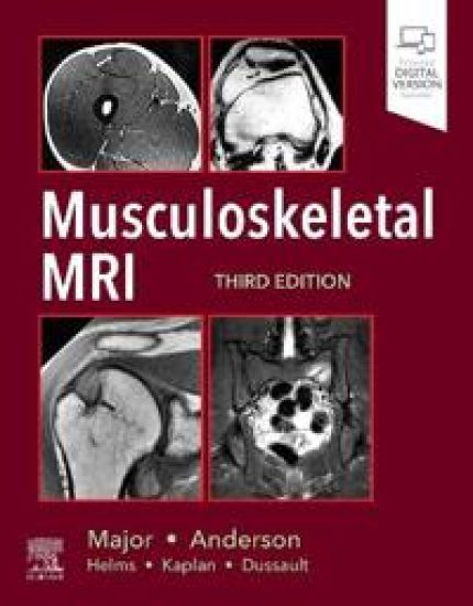
Musculoskeletal MRI
Nancy M. Major; Mark W. Anderson
Elsevier - Health Sciences Division
2019
sidottu
Halvin toimitettuna 168,90 €
Ideal for residents, practicing radiologists, and fellows alike, this updated reference offers easy-to-understand guidance on how to approach musculoskeletal MRI and recognize abnormalities. Concise, to-the-point text covers MRI for the entire musculoskeletal system, presented in a highly templated format. Thoroughly revised and enhanced with full-color artwork throughout, this resource provides just the information you need to perform and interpret quality musculoskeletal MRI. Includes the latest protocols, practical advice, tips, and pearls for diagnosing conditions impacting the temporomandibular joint, shoulder, elbow, wrist/hand, spine, hips and pelvis, knee, and foot and ankle. Follows a quick-reference format throughout, beginning with basic technical information on how to obtain a quality examination, followed by a discussion of the normal appearance and the abnormal appearance for each small unit that composes a joint. Depicts both normal and abnormal anatomy, as well as disease progression, through more than 600 detailed, high-quality images, most of which are new to this edition. Features key information boxes throughout for a quick review of pertinent material. Expert ConsultT eBook version included with purchase. This enhanced eBook experience allows you to search all of the text, figures, and references from the book on a variety of devices.
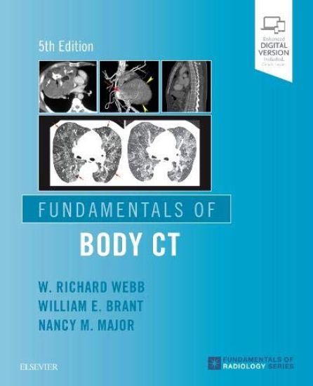
Fundamentals of Body CT
W Richard Webb; Wiliam E. Brant; Nancy M. Major
Elsevier - Health Sciences Division
2019
nidottu
From recent advances in helical CT techniques to new developments in lung cancer screening to optimized CT techniques in musculoskeletal diagnosis, Fundamentals of Body CT, 5th Edition, covers the essential information you need to know to effectively perform and interpret CT scans. Step-by-step instructions for all current CT techniques help you quickly understand each procedure and review key steps. Comprehensive and easy to digest, this introduction to body CT is an essential resource for radiology residents, practicing radiologists, and medical students. Features many new topics, discussions of additional diseases, and new, high-quality images from cover to cover, including updated descriptions and illustrations of normal anatomy and incidental findings. Allows you to quickly compare diagnoses with a survey of major CT findings for a variety of common diseases?with an emphasis on those findings that help to differentiate one condition from another. Reviews the spiral/helical CT protocols currently used for the diagnosis of chest, abdominal, and musculoskeletal abnormalities, including high-resolution CT, lung nodule assessment and lung cancer screening, CT pulmonary embolism diagnosis, CT enterography, CT enteroclysis, CT colonography, and optimizing CT techniques in musculoskeletal diagnosis. Brings you up to date with recent advances in chest CT, including the classification of adenocarcinoma, evaluation of lung nodules, lung cancer screening (including Lung-RADS) and staging, and the classification and diagnosis of interstitial lung diseases using high-resolution CT Covers new developments in abdominal CT such as the Liver Imaging Reporting and Data System (Li-RADS) for imaging and reporting small hepatocellular carcinoma, reviews of the Atlanta Classification of Acute Pancreatitis, and an improved description of CT findings of histologic subtypes of renal cell carcinoma. Includes new discussions of the diagnosis of musculoskeletal abnormalities detected on chest and abdominal CT scans obtained for non-musculoskeletal indications. Contains updated disease classifications, including those for pulmonary adenocarcinoma, diffuse lung diseases, and pancreatic lesions. Enhanced eBook version included with purchase. Your enhanced eBook allows you to access all of the text, figures, and references from the book on a variety of devices.

Fundamentals of Body CT
W. Richard Webb; Wiliam E. Brant; Nancy M. Major
Elsevier India
2014
nidottu
Approx. 386 pages
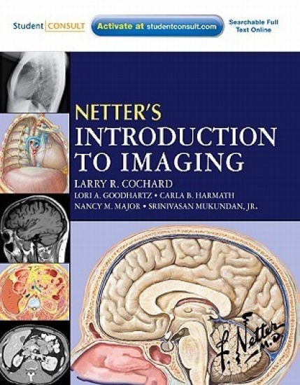
Netter's Introduction to Imaging
Larry R. Cochard; Lori A Goodhartz; Carla Harmath; Nancy M. Major; Srinivasan Mukundan
W B Saunders Co Ltd
2011
nidottu
Netter's Introduction to Imaging, by Larry R. Cochard, PhD, Lori A Goodhartz, MD Carla B, Harmath, MD, Nancy M. Major MD, and Srinivasan Mukundan, JR, MD, makes interpreting normal and abnormal X-ray, CT, and MR images easy by correlating them with crystal-clear Netter illustrations. You'll learn to recognize anatomical relationships in images and apply them to a variety of examples of pathology throughout the body, including the imaging of masses, air, or blood in organs and spaces...fractures, thickening, constriction, and compression...and more. You can also access the book from any computer at studentconsult.com. It's an ideal introduction to diagnostic imaging! Visualize anatomical structures and relationships with perfect clarity with the aid of vivid, colorful Netter artwork. The coloring, texture, and idealized emphasis help you interpret relationships between structures and compartments as seen in cross section and in X-rays, CT, and MRI. Develop your ability to better identify pathologies by viewing normal healthy anatomical images and abnormal images. Comparative images reinforce your basic understanding of what normal tissues and anatomy look like and serve as a guide in recognizing disease patterns and processes: atypically large or small organs and compartments, masses, air, or blood in organs and spaces, fractures, thickening, constriction, compression, and more. Understand the principles that underlie X-ray, CT, MR, ultrasound, and nuclear medicine imaging, the use of contrast and angiography. Understand how radiologists apply systematic search strategies in imaging studies of each region of the body. Access the complete text and images online at studentconsult.com
This lavishly illustrated book is a perfect introduction to the exciting field of radiology. A lively, easy-to-read writing style explores the diagnostic imaging issues associated with each body region. Extensive tables, color-coded key points, and detailed drawings make finding essential information a snap. Case studies and high- resolution images demonstrate radiologic principles and characteristic findings for a wide range of disease entities. All imaging modalities are covered including fluoroscopy, CT, MR, nuclear medicine, and more. An ideal text for anyone interested in diagnostic imaging, A Practical Approach to Radiology not only gives you the information you need, it brings it to life. Features a full-color, templated format, making pertinent information easy to digest and recall. Includes color-coded tables that discuss differential diagnoses and indications for types of exams. Uses key points to list the findings on an x-ray as well as outlining search patterns. Explores the latest, cutting-edge technologies in the field, including spiral CT and 3-D reconstruction. Contains over 800 images to demonstrate important points.