Kirjojen hintavertailu – 12 903 725 kirjaa ja 27 kauppaa

Kirjailija

Parvez Sheik

Kirjat ja teokset yhdessä paikassa: 3 kirjaa, julkaisuja vuosilta 2011–2017, suosituimpiin kuuluu Atlas of Human Anatomy on MRI. Vertaile teosten hintoja ja tarkista saatavuus suomalaisista kirjakaupoista.

3 kirjaa

Kirjojen julkaisuvuodet: 2011–2017.

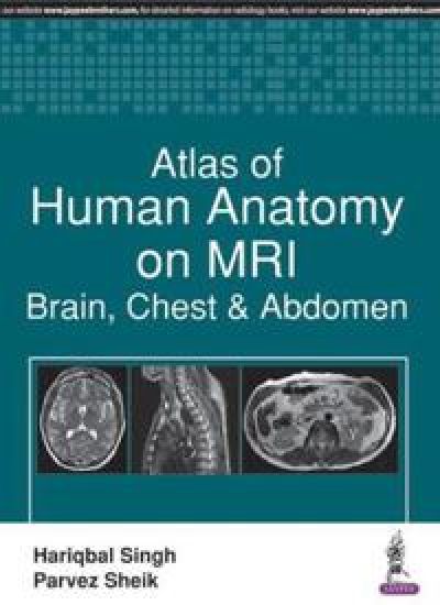
Atlas of Human Anatomy on MRI

Atlas of Human Anatomy on MRI

Hariqbal Singh; Parvez Sheik

Jaypee Brothers Medical Publishers
2017
nidottu

Halvin toimitettuna 70,90 €

This book is a concise overview of MRI (magnetic resonance imaging) for brain, chest and abdominal disorders covering the very latest technologies and developments in the field. Beginning with an introduction to anatomy of these body systems, the following sections cover MR cholangiopancreatography, MRI of the female and male pelvis, and MR angiography. The atlas is enhanced by high quality MR images and tables with detailed descriptions to help clinicians understand complex anatomy. The comprehensive appendix provides a glossary of MRI terms and radiology measurement tables. Key Points Concise overview of MRI for brain, chest and abdomenFeatures sections on MR cholangiopancreatography, MRI of the pelvis, and MR angiographyComprehensive appendix provides glossary of terms and radiology measurement tablesIncludes high quality MR images and tables illustrating complex anatomy
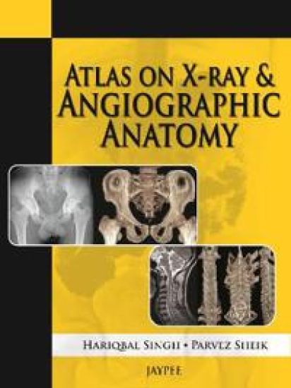
Atlas on X-Ray and Angiographic Anatomy

Atlas on X-Ray and Angiographic Anatomy

Hariqbal Singh; Parvez Sheik

Jaypee Brothers Medical Publishers
2013
nidottu
This atlas presents trainees with numerous X-ray and angiographic images to gain a thorough understanding of normal radiographic anatomy in order to make an accurate diagnosis of underlying pathology. Presented in an easy to read format, the book covers radiological procedures, ossification centres, X-ray production, digital subtraction angiography, and computed and digital radiography, in the different anatomical sections of the body. This practical guide includes nearly 240 clearly labelled images, illustrations and tables, with detailed descriptions, to assist learning. Key points Atlas of X-ray and angiographic images to help trainees understand normal radiographic anatomy and diagnose underlying pathologyEasy to read formatCovers different imaging techniques for all areas of the bodyIncludes nearly 240 images, illustrations and tables with detailed descriptions