
Kirjojen hintavertailu – 12 903 725 kirjaa ja 27 kauppaa

Kirjailija

Sherief Garrana

Kirjat ja teokset yhdessä paikassa: 2 kirjaa, julkaisuja vuosilta 2023–2025, suosituimpiin kuuluu Imaging Anatomy: Chest, Abdomen, Pelvis. Vertaile teosten hintoja ja tarkista saatavuus suomalaisista kirjakaupoista.

2 kirjaa

Kirjojen julkaisuvuodet: 2023–2025.

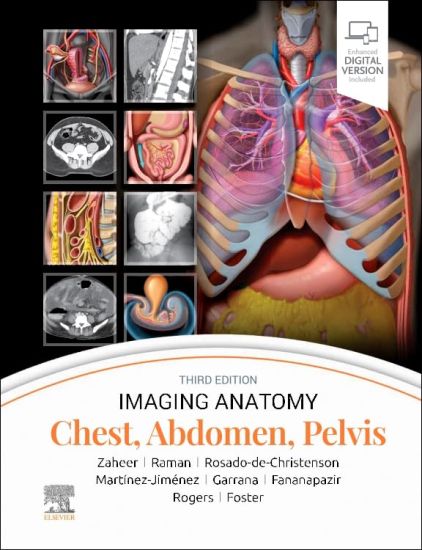
Imaging Anatomy: Chest, Abdomen, Pelvis

Imaging Anatomy: Chest, Abdomen, Pelvis

Siva P. Raman; Melissa L. Rosado-de-Christenson; Atif Zaheer; Santiago Martínez-Jiménez; Ghaneh Fananapazir; Sherief Garrana; Douglas Rogers; Bryan R. Foster

Elsevier Health Sciences
2023
sidottu
This richly illustrated and superbly organized text/atlas is an excellent point-of-care resource for practitioners at all levels of experience and training. Written by global leaders in the field, Imaging Anatomy: Chest, Abdomen, Pelvis, third edition, contains specifics about radiographic, multiplanar, high-resolution, and cross-sectional body imaging along with thousands of relevant examples to give busy clinicians quick answers to imaging anatomy questions. This must-have reference employs a templated, highly formatted design; concise, bulleted text; and state-of-the-art images throughout that identify characteristic normal imaging findings and anatomic variants in each anatomic area, offering a unique opportunity to master the fundamentals of normal anatomy and accurately and efficiently recognize pathologic conditions. Contains nearly 2,800 print and online-only images, including all relevant imaging modalities, 3D reconstructions, and detailed, high-resolution medical drawings that together illustrate the fine points of imaging anatomy Reflects new understandings of anatomy due to ongoing anatomic research as well as new, advanced imaging techniques Offers new content on the anatomic basis for thoracic developmental abnormalities, anatomic variants of systemic and pulmonary vasculature, and the PI-RADS system and clinical implications of MR for prostate cancer Contains new and updated images of the chest wall musculature with CT and MR examples; abdominal imaging best practices, including the application of body MR in the abdomen and pelvis; and the different modalities used for GU/GYN imaging, specifically retrograde urethrography and MR for specific disease diagnosis Depicts common anatomic variants and covers the common pathological processes that manifest with alterations of normal anatomic landmarks Features representative pathologic examples to highlight the effect of disease on human anatomy Presents essential text in an easy-to-digest, bulleted format, enabling imaging specialists to find quick answers to anatomy questions encountered in daily practice Includes an eBook version that enables you to access all text, figures, and references with the ability to search, customize your content, make notes and highlights, and have content read aloud
Specialty Imaging: HRCT of the Lung

Specialty Imaging: HRCT of the Lung

Santiago Martínez-Jiménez; Melissa L. Rosado-de-Christenson; Sherief Garrana

Elsevier Health Sciences
2025
sidottu

Halvin toimitettuna 264,90 €

Part of the highly regarded Specialty Imaging series, HRCT of the Lung, third edition, reflects the many recent changes in HRCT diagnostic interpretation. An easy-to-read bulleted format and thousands of state-of-the-art imaging examples guide you step by step through every aspect of thin-section CT and HRCT in the evaluation of patients with suspected lung disease. This book is an ideal resource for radiologists and internal medicine specialists who need an easily accessible tool to help them understand the indications, strengths, and limitations of HRCT in their practice. Helps you identify and characterize critical HRCT findings and gain a solid understanding of cross-sectional imaging anatomy of the lung-crucial skills for formulating diagnoses and appropriate differential diagnoses Delivers details on anatomy-based exploration as well as nodules and micronodules, cysts and pseudocysts, reticulation and honeycombing, mosaic attenuation, ground-glass opacities, and interlobular septal thickening Uses a time-saving, bulleted format that distills essential information for fast and easy comprehension Includes new chapters on post-COVID-19 findings, respiratory syncytial virus, and e-cigarette and vaping product use-associated lung injury (EVALI), as well as content on new morphologic patterns for pulmonary fibrosis, the new classification for hypersensitivity pneumonia, and more Provides direction for determining common, less common, and rare diagnoses based on morphologic features, distribution of abnormalities, and/or histologic patterns Features superb illustrations with comprehensive captions that display both typical and variant findings on HRCT scans Offers new references, new images, and new histopathologic correlations throughout Includes an eBook version that allows you to access everything in this print version as well as additional images, text, and references, with the ability to search, customize your content, make notes and highlights, and have content read aloud; additional digital ancillary content may publish up to 6 weeks following the publication date