Kirjojen hintavertailu – 12 903 725 kirjaa ja 27 kauppaa

Kirjailija

Siva P. Raman

Kirjat ja teokset yhdessä paikassa: 4 kirjaa, julkaisuja vuosilta 2021–2025, suosituimpiin kuuluu Imaging Anatomy: Chest, Abdomen, Pelvis. Vertaile teosten hintoja ja tarkista saatavuus suomalaisista kirjakaupoista.

4 kirjaa

Kirjojen julkaisuvuodet: 2021–2025.

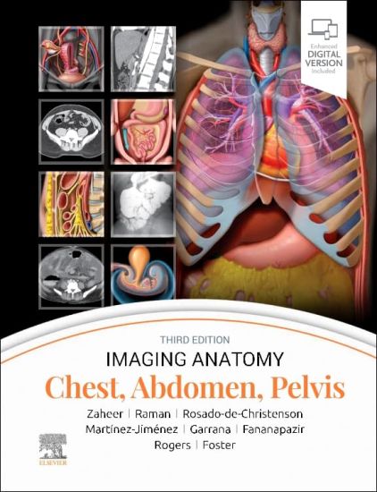
Imaging Anatomy: Chest, Abdomen, Pelvis

Imaging Anatomy: Chest, Abdomen, Pelvis

Siva P. Raman; Melissa L. Rosado-de-Christenson; Atif Zaheer; Santiago Martínez-Jiménez; Ghaneh Fananapazir; Sherief Garrana; Douglas Rogers; Bryan R. Foster

Elsevier Health Sciences
2023
sidottu
This richly illustrated and superbly organized text/atlas is an excellent point-of-care resource for practitioners at all levels of experience and training. Written by global leaders in the field, Imaging Anatomy: Chest, Abdomen, Pelvis, third edition, contains specifics about radiographic, multiplanar, high-resolution, and cross-sectional body imaging along with thousands of relevant examples to give busy clinicians quick answers to imaging anatomy questions. This must-have reference employs a templated, highly formatted design; concise, bulleted text; and state-of-the-art images throughout that identify characteristic normal imaging findings and anatomic variants in each anatomic area, offering a unique opportunity to master the fundamentals of normal anatomy and accurately and efficiently recognize pathologic conditions. Contains nearly 2,800 print and online-only images, including all relevant imaging modalities, 3D reconstructions, and detailed, high-resolution medical drawings that together illustrate the fine points of imaging anatomy Reflects new understandings of anatomy due to ongoing anatomic research as well as new, advanced imaging techniques Offers new content on the anatomic basis for thoracic developmental abnormalities, anatomic variants of systemic and pulmonary vasculature, and the PI-RADS system and clinical implications of MR for prostate cancer Contains new and updated images of the chest wall musculature with CT and MR examples; abdominal imaging best practices, including the application of body MR in the abdomen and pelvis; and the different modalities used for GU/GYN imaging, specifically retrograde urethrography and MR for specific disease diagnosis Depicts common anatomic variants and covers the common pathological processes that manifest with alterations of normal anatomic landmarks Features representative pathologic examples to highlight the effect of disease on human anatomy Presents essential text in an easy-to-digest, bulleted format, enabling imaging specialists to find quick answers to anatomy questions encountered in daily practice Includes an eBook version that enables you to access all text, figures, and references with the ability to search, customize your content, make notes and highlights, and have content read aloud
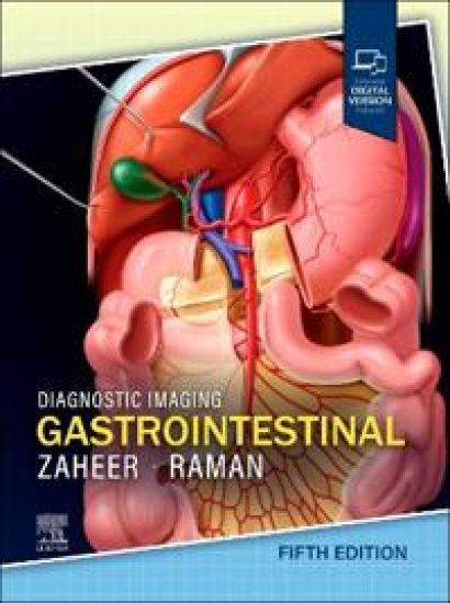
Diagnostic Imaging: Gastrointestinal

Diagnostic Imaging: Gastrointestinal

Atif Zaheer; Siva P. Raman

Elsevier Health Sciences
2025
sidottu
Covering the entire spectrum of this fast-changing field, the fifth edition of Diagnostic Imaging: Gastrointestinal is an invaluable resource for GI radiologists, general radiologists, and trainees?anyone who requires an easily accessible, highly visual reference on today’s GI imaging. Drs. Atif Zaheer, Siva P. Raman, and their team of highly regarded experts provide updated information on recent advances in technology and the understanding of GI diseases and disorders to help you make informed decisions at the point of care. The text is image-rich, with succinct bullets that quickly convey details, and includes the latest literature references, making it a useful learning tool as well as a handy reference for daily practice. Serves as a one-stop resource for key concepts and information on GI imaging, including a wealth of new material and content updates throughout Covers all aspects of GI imaging, including pathophysiology, imaging findings, and disease management options, such as the radiologist’s role in evaluating patients for bariatric surgery, antireflux procedures, esophageal and bowel resections, and more Features 3,300+ high-quality print images (with an additional 2,100+ images in the complimentary eBook), including radiology images, detailed medical illustrations, and clinical photographs, as well as histology and gross pathology images Features updates from cover to cover, including new information on rectal MR, pelvic floor MR, and MR elastography; updated CT protocols; updated differential diagnoses; new cases and images; and new staging details and diagrams Contains a variety of new chapters, including Approach to Incidental Lymph Nodes on CT/MR: Summary of 2013 ACR Consensus Statement; Approach to Management of Incidental Splenic Lesions: Summary of 2013 ACR Consensus Statement; Management of Incidental Gallbladder and Biliary Findings: Summary of Consensus Statements; Management of Incidental Liver Lesions; Management of Pancreatic Cysts (with new guidelines); Summary of C-RADS for CT Colonography; and Summary of LI-RADS for CT and MR Imaging Uses bulleted, succinct text and highly templated chapters for quick comprehension of essential information at the point of care Includes an eBook that allows you access to everything in the print version as well as additional images, text, and references, with the ability to search, customize your content, make notes and highlights, and have content read aloud
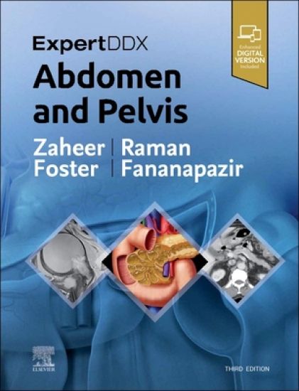
ExpertDDx: Abdomen and Pelvis

ExpertDDx: Abdomen and Pelvis

Atif Zaheer; Siva P. Raman; Bryan R. Foster; Ghaneh Fananapazir

Elsevier - Health Sciences Division
2022
sidottu
Highly practical and user-friendly, ExpertDDx: Abdomen and Pelvis, third edition, helps you reach accurate, clinically useful differential diagnoses in your everyday practice. It presents the most useful differential diagnoses for each region of the abdomen and pelvis, grouped according to anatomic location, generic imaging findings, modality-specific findings, or clinical-based indications. Each differential diagnosis includes several high-quality, succinctly annotated images; a list of diagnostic possibilities sorted as common, less common, and rare but important; and brief, bulleted text offering helpful diagnostic clues. It's an excellent resource for subspecialty abdominal imagers as well as general radiologists and trainees, providing invaluable assistance in reaching logical, on-target differential diagnoses based on key imaging findings and clinical details. Covers 175 of the most common diagnostic challenges in abdominal and pelvic imaging, enhanced by more than 2,100 radiologic images, full-color illustrations, clinical and histologic photographs, and gross pathology images Provides a quick review of the salient features of each entity, differentiating features from other similar-appearing abnormalities Includes new chapters on hematuria, flank pain, acute scrotal pain, and seminal vesicle Adds greater focus to advancing prostate imaging methods with expanded content on lesions in the peripheral zone and lesions in the transition zone, as well as new coverage of transplant imaging Contains updates to numerous classifications, including LI-RADS for liver, O-RADS for ovarian masses, and the Tanaka classification for pancreatic cysts Features new MR examples and MR-specific diagnoses throughout, plus new differentials for contrast-enhanced ultrasound findings related to liver and kidney lesions Includes the enhanced eBook version, which allows you to search all text, figures, and references on a variety of devices
Diagnostic Imaging: Gastrointestinal

Diagnostic Imaging: Gastrointestinal

Atif Zaheer; Siva P. Raman

Elsevier - Health Sciences Division
2021
sidottu
Covering the entire spectrum of this fast-changing field, Diagnostic Imaging: Gastrointestinal, fourth edition, is an invaluable resource for gastrointestinal radiologists, general radiologists, and trainees-anyone who requires an easily accessible, highly visual reference on today's GI imaging. Drs. Siva P. Raman, Atif Zaheer, and their team of highly regarded experts provide up-to-date information on recent advances in technology and the understanding of GI diseases and disorders to help you make informed decisions at the point of care. The text is lavishly illustrated, delineated, and referenced, making it a useful learning tool as well as a handy reference for daily practice. Serves as a one-stop resource for key concepts and information on gastrointestinal imaging, including a wealth of new material and content updates throughout Features more than 2,900 illustrations (multiplanar CT, sonography, MR, and PET/CT; clinical photos; radiologic images; histologic images; H&E stains; and full-color illustrations) as well as an additional 3K digital-only images and new video clips Features updates from cover to cover including new information on MRI imaging of rectal cancer, iron quantification, and MRI protocols; new cases and images, and new staging details and diagrams Contains new chapters on treatment response criteria for systemic conditions (RECIST, irRECIST, etc.), dual energy CT for pancreas, vascular abnormalities, MR elastography of the liver, pretransplant liver evaluation, and more Covers all aspects of GI imaging, including pathophysiology, imaging findings, and disease management options such as the radiologist's role in evaluating patients for bariatric surgery, antireflux procedures, esophageal and bowel resections, and more Uses bulleted, succinct text and highly templated chapters for quick comprehension of essential information at the point of care Enhanced eBook version included with purchase. Your enhanced eBook allows you to access all of the text, figures, and references from the book on a variety of devices