
Kirjojen hintavertailu – 12 903 725 kirjaa ja 27 kauppaa

Kirjailija

Srinivasan Mukundan

Kirjat ja teokset yhdessä paikassa: 2 kirjaa, julkaisuja vuosilta 2011–2014, suosituimpiin kuuluu Netter's Correlative Imaging: Neuroanatomy. Vertaile teosten hintoja ja tarkista saatavuus suomalaisista kirjakaupoista.

2 kirjaa

Kirjojen julkaisuvuodet: 2011–2014.

Netter's Correlative Imaging: Neuroanatomy

Netter's Correlative Imaging: Neuroanatomy

Thomas C. Lee; Srinivasan Mukundan

W B Saunders Co Ltd
2014
sidottu
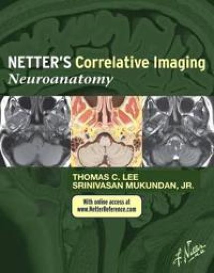
Interpret the complexities of neuroanatomy like never before with the unparalleled coverage and expert guidance from Drs. Srinivasan Mukundan and Thomas C. Lee in this outstanding volume of the Netter's Correlative Imaging series. Beautiful and instructive Netter paintings and illustrated cross-sections created in the Netter style are presented side by side high-quality patient images and key anatomic descriptions to help you envision and review intricate neuroanatomy. View the brain, spinal cord, and cranial nerves, as well as head and neck anatomy through modern imaging techniques in a variety of planes, complemented with a detailed illustration of each slice done in the instructional and aesthetic Netter style. Find anatomical landmarks quickly and easily through comprehensive labeling and concise text highlighting key points related to the illustration and image pairings. Correlate patient data to idealized normal anatomy, always in the same view with the same labeling system.
Netter's Introduction to Imaging

Netter's Introduction to Imaging

Larry R. Cochard; Lori A Goodhartz; Carla Harmath; Nancy M. Major; Srinivasan Mukundan

W B Saunders Co Ltd
2011
nidottu
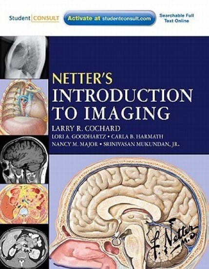
Netter's Introduction to Imaging, by Larry R. Cochard, PhD, Lori A Goodhartz, MD Carla B, Harmath, MD, Nancy M. Major MD, and Srinivasan Mukundan, JR, MD, makes interpreting normal and abnormal X-ray, CT, and MR images easy by correlating them with crystal-clear Netter illustrations. You'll learn to recognize anatomical relationships in images and apply them to a variety of examples of pathology throughout the body, including the imaging of masses, air, or blood in organs and spaces...fractures, thickening, constriction, and compression...and more. You can also access the book from any computer at studentconsult.com. It's an ideal introduction to diagnostic imaging! Visualize anatomical structures and relationships with perfect clarity with the aid of vivid, colorful Netter artwork. The coloring, texture, and idealized emphasis help you interpret relationships between structures and compartments as seen in cross section and in X-rays, CT, and MRI. Develop your ability to better identify pathologies by viewing normal healthy anatomical images and abnormal images. Comparative images reinforce your basic understanding of what normal tissues and anatomy look like and serve as a guide in recognizing disease patterns and processes: atypically large or small organs and compartments, masses, air, or blood in organs and spaces, fractures, thickening, constriction, compression, and more. Understand the principles that underlie X-ray, CT, MR, ultrasound, and nuclear medicine imaging, the use of contrast and angiography. Understand how radiologists apply systematic search strategies in imaging studies of each region of the body. Access the complete text and images online at studentconsult.com