
Kirjojen hintavertailu – 12 859 829 kirjaa ja 27 kauppaa

Kirjailija

Sujal Desai

Kirjat ja teokset yhdessä paikassa: 2 kirjaa, julkaisuja vuosilta 2007–2021, suosituimpiin kuuluu Pulmonary Imaging. Vertaile teosten hintoja ja tarkista saatavuus suomalaisista kirjakaupoista.

2 kirjaa

Kirjojen julkaisuvuodet: 2007–2021.

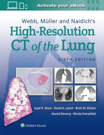
Webb, Müller and Naidich's High-Resolution CT of the Lung

Webb, Müller and Naidich's High-Resolution CT of the Lung

Sujal Desai; David Lynch; Brett M Elicker; Anand Devaraj; Nicola Sverzellati

Wolters Kluwer Health
2021
sidottu
Selected as a Doody’s Essential Title for 2025?! Over the past 30 years high-resolution CT (HRCT) has matured to become an integral part of the multidisciplinary evaluation in diffuse lung disease. In this regard, Webb, Muller and Naidich’s High-Resolution CT of the Lung, 6th Edition, is a ‘gold standard’ reference that aims to keep radiologists and pulmonologists alike at the cutting edge of the ever-evolving field of thoracic imaging. The new US-European author team continues the tradition of excellence which readers have come to expect while the underlying layout and ethos — established by the ‘founding’ author team — remain. The new edition aims to brings readers up to date not only with recent advances but also with the important conceptual changes in thinking in various fields of thoracic imaging. Also featured in this updated edition is authoritative guidance on HRCT findings and differential diagnosis, as well as the characteristics of the common lung diseases assessed using HRCT, all enhanced by a multitude of new images and updated content throughout. Covers the physical and anatomic principles governing HRCT imaging and the importance of the ‘pattern-based’ approach to HRCT interpretation. Updates to chapters on interstitial lung diseases and airways diseases & connective tissue diseases reflect the dramatic changes in thinking in these fields. Includes new and improved features including key HRCT findings in separate tables in relevant sections, where relevant, and new and exciting topics including artificial intelligence and machine learning. [Reword to: Includes improved features such as key HRCT findings in separate tables, and new topics including artificial intelligence and machine learning. ?] Contains full-color illustrations of histologic findings in lung disease linked with HRCT manifestations. Provides diagnostic algorithms, an illustrated quick reference guide at the front of the text, and helpful tables and line drawings. Enrich Your eBook Reading Experience Read directly on your preferred device(s), such as computer, tablet, or smartphone. Easily convert to audiobook, powering your content with natural language text-to-speech.
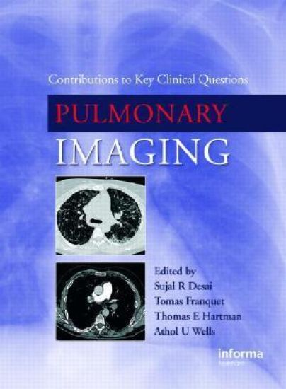
Pulmonary Imaging

Pulmonary Imaging

Sujal Desai; Tomas Franquet; Thomas E. Hartman; Athol Wells

CRC Press
2007
sidottu
Unlike other texts of imaging for clinicians, Pulmonary Imaging seeks not to teach the physician or surgeon about the radiologic signs of pulmonary disease but rather inform about the value of the various imaging tests in different clinical scenarios. Accordingly, each chapter is written by a clinician and radiologist: the basic ethos is that the clinician identifies the common problem-areas and questions faced in a given field of pulmonology and that the radiologist attempts to critically define the role, if any, of the different imaging studies. Pulmonary Imaging will be valued by physicians and surgeons who regularly investigate and manage patients with pulmonary disease. There is little doubt that imaging tests are vital in the investigation of respiratory disease. However, because of the staggering advances in imaging technology, the practicing physician or surgeon is often faced with a wide range of potential radiologic tests. Moreover, it is fair to state that the evidence-base for the utility of some tests in specific clinical scenarios sometimes lags behind the enthusiasm and adoption in practice. Indeed, it has become apparent to many thoracic radiologists that, in certain clinical settings, the more sophisticated radiologic techniques (e.g. multidetector row CT) probably have little to offer over the more traditional modalities, such as plain radiography. The main aim of Pulmonary Imaging: Contributions to Key Clinical Questions is to inform the reader about the value of various imaging tests in different clinical scenarios, based on the available evidence. Each chapter is co-authored by a physician and radiologist with the former defining the key clinical questions which arise in a specific setting and the radiologist outlining the role of imaging tests, if any, in answering these questions. It is hoped that this critical approach to radiologic tests will appeal not only to physicians and surgeons but also to radiologists who must regularly manage patients with respiratory disease.