Kirjojen hintavertailu – 12 903 725 kirjaa ja 27 kauppaa

Kirjailija

Wolfgang Seeger

Kirjat ja teokset yhdessä paikassa: 12 kirjaa, julkaisuja vuosilta 1968–2020, suosituimpiin kuuluu Endoscopic and microsurgical anatomy of the upper basal cisterns. Vertaile teosten hintoja ja tarkista saatavuus suomalaisista kirjakaupoista.

12 kirjaa

Kirjojen julkaisuvuodet: 1968–2020.

Evolution of the Central Nervous System of Craniata and Homo

Evolution of the Central Nervous System of Craniata and Homo

Wolfgang Seeger

Springer Nature Switzerland AG
2020
nidottu
The main focus of this book is on providing students, neurosurgery trainees, certified neurosurgeons and colleagues in neighbouring disciplines essential information on the evolution of the central nervous system (CNS) of craniata and homo. Therefore the book is divided in three parts: Part I is describing the evolution of CNS of craniata (starting 800 million of years ago). Part II is explaining in detail the exceptional position of the human encephalon. Part III is discussing maturity and immaturity of all parts of CNS of craniatas and the consequences concerning further development of brain structure and psychological functions. In all parts anatomical fundamentals are presented in the form of didactic and self-explanatory illustrations.

Evolution of the Central Nervous System of Craniata and Homo

Evolution of the Central Nervous System of Craniata and Homo

Wolfgang Seeger

Springer Nature Switzerland AG
2019
sidottu
The main focus of this book is on providing students, neurosurgery trainees, certified neurosurgeons and colleagues in neighbouring disciplines essential information on the evolution of the central nervous system (CNS) of craniata and homo. Therefore the book is divided in three parts: Part I is describing the evolution of CNS of craniata (starting 800 million of years ago). Part II is explaining in detail the exceptional position of the human encephalon. Part III is discussing maturity and immaturity of all parts of CNS of craniatas and the consequences concerning further development of brain structure and psychological functions. In all parts anatomical fundamentals are presented in the form of didactic and self-explanatory illustrations.
Anatomical Basis of Cranial Neurosurgery

Anatomical Basis of Cranial Neurosurgery

Wolfgang Seeger; Josef Zentner

Springer Nature Switzerland AG
2019
nidottu
This didactic book clearly and systematically describes the anatomical-surgical fundamentals of cranial neurosurgery, relating them to norm variants, imaging modalities and interdisciplinary aspects. All illustrations, hand drawn in ink by the first author, are simple and self-explanatory. The book reflects the first author’s lifetime experience as an academic neurosurgeon and teacher, as well as the second author’s theoretical and practical knowledge of neurosurgical subspecialties such as epilepsy surgery. In addition to its core audience in neurosurgery, it provides all connected disciplines, in particular neuroradiology, neurology, neuropathology, ENT surgery, maxillofacial surgery and eye surgery, with unique anatomical insights into the neurosurgeon’s perspective.
Microanatomical Aspects for Neurosurgeons and Neuroradiologists
Modern diagnostic imaging and operative approaches have witnessed significant improvements in our times. Computerassisted methods are in use in all microsurgical fields. Neuronavigation, novel stereotactic methods, endoscopic procedures, magnetic resonance imaging, ultrasound and the progress in pre- and intraoperative epilepsy diagnostics must be mentioned in particular in this connection. However, the insights of neuroanatomy and neurophysiology have not become obsolete thereby, on the contrary: such knowledge is imperative and a prerequisite for all neurosurgeons, nowadays more than ever before. Otherwise, excellent modern approaches are liable to fall into discredit if microanatomical aspects are neglected. The goal of this book is two-fold: first, to guide the resident towards a fruitful application of anatomical basics in visualizing and operative techniques. Second, to draw attention to as many anatomical norm variants as possible to forestall complications during surgery. Standard methods, such as the pterional approach, often confront the surgeon with a range of anatomical variants.
Anatomical Basis of Cranial Neurosurgery

Anatomical Basis of Cranial Neurosurgery

Wolfgang Seeger; Josef Zentner

Springer International Publishing AG
2018
sidottu
This didactic book clearly and systematically describes the anatomical-surgical fundamentals of cranial neurosurgery, relating them to norm variants, imaging modalities and interdisciplinary aspects. All illustrations, hand drawn in ink by the first author, are simple and self-explanatory. The book reflects the first author’s lifetime experience as an academic neurosurgeon and teacher, as well as the second author’s theoretical and practical knowledge of neurosurgical subspecialties such as epilepsy surgery. In addition to its core audience in neurosurgery, it provides all connected disciplines, in particular neuroradiology, neurology, neuropathology, ENT surgery, maxillofacial surgery and eye surgery, with unique anatomical insights into the neurosurgeon’s perspective.
Endoscopic and microsurgical anatomy of the cranial base
Neuroendoscopic transnasal surgical approaches have become increasingly common for some time, and have started to replace microsurgical techniques in pituitary surgery. Endoscopic transnasal approaches have also been recently used in some neurosurgical centres to reach pre- and retrosellar areas and targets localized in the basal cisterns. One major factor is the localization of the carotid artery between the siphon and aperture externa of the carotid canal at the base of the petrous bone. The following areas are especially critical: 1. the anterior siphon area 2.the area between the bottom of the sphenoid sinus and the base of Processus pte- goideus 3. the proximity of the carotid canal to the Tuba Eustachii, the labyrinth, Bulbus su- rior of the internal jugular vein and the facial nerve. Some segments of the course of the carotid artery are well known but they have rarely been surgical target areas using transnasal approaches to date. This has changed since the introduction of modern imaging techniques, especially neuronavigation. It has become possible to identify and localize structures of the skull and extra- and intracranial structures in any desired plane.
Standard Variants of the Skull and Brain

Standard Variants of the Skull and Brain

Wolfgang Seeger

Springer Verlag GmbH
2014
nidottu
The author describes in his unique style the anatomical variants of the brain and skull. This atlas is a continuation of his last work on "Neuronavigation and Neuroanatomy". Most anatomical reference volumes show a large number of common and rare variations. This atlas concentrates on well known and little known variants which are especially important for the clinicians, in particular the neurosurgeons and the radiologists. The variants have been grouped after areas of trepanation. The author presents also a number of so far unknown variants gathered from his personal theoretical and clinical experience of 50 years. Exact knowledge of anatomical variations which the surgeon may encounter helps to plan operations and to avoid unexpected complications. Variants of no clinical relevance, even rather common ones, have not been included.
Compressive Optic Nerve Lesions at the Optic Canal

Compressive Optic Nerve Lesions at the Optic Canal

Renate Unsöld; Wolfgang Seeger

Springer-Verlag Berlin and Heidelberg GmbH Co. K
2011
nidottu
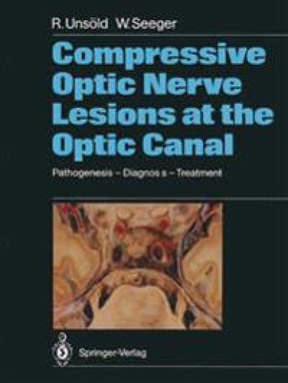
The optic canal, in particular its intracranial end, represents a "locus minoris resistentiae" for optic nerve compression in a variety of pathologic conditions. The intracranial optic nerve shares the limited space within this narrow passage with the carotid and ophthalmic artery, all being surrounded by bone and rigid dura. Any pathological condition going along with an increase of soft tissue volume, such as in optic nerve sheath tumors, parasellar neoplasms, dolichoectasia of the carotid and/ or ophthalmic artery, hematomas, etc. , or reduction of the lumen of the bony optic canal by hyperpneumatization of the sphenoid sinus, hyperostosis or developmental abnormalities must act as a space-occupying lesion causing optic nerve compression either by pressing the nerve against the vessel or the neighboring dura or bone. The spectrum of clinical signs and symptoms of optic nerve compression in this area is rather wide and includes acute as well as slowly progressive visual loss and all kinds of visual field defects in the presence of a normal disk, papilledema, pri- mary optic atrophy or cavernous optic atrophy mimicking var- ious clinical disease entities such as retrobulbar optic neuritis, anterior and posterior ischemic optic neuropathy, soft glaucoma and others. Some of the lesions causing optic nerve compression in this area are rather small and need to be visualized or excluded by thin section CT such as pneumosinus dilatans of the sphenoid bone, dolichoectasia of the internal carotid artery, small men- ingiomas around the optic foramen and others.
Endoscopic and microsurgical anatomy of the upper basal cisterns
This detailed atlas illustrates the anatomical structures of the upper basal cisterns, their topography and relationship to other intra- and extradural structures. The author expands his well-established efforts to convey his outstanding neuroanatomical knowledge to the basal cisterns. His famous anatomical drawings are based upon anatomical preparations, cadaver dissections and intraoperative pictures, in order to point out important aspects concerning microsurgical and endoscopic approaches to these parts of the brain.
Endoscopic and microsurgical anatomy of the cranial base
Neuroendoscopic transnasal surgical approaches have become increasingly common for some time, and have started to replace microsurgical techniques in pituitary surgery. Endoscopic transnasal approaches have also been recently used in some neurosurgical centres to reach pre- and retrosellar areas and targets localized in the basal cisterns. One major factor is the localization of the carotid artery between the siphon and aperture externa of the carotid canal at the base of the petrous bone. The following areas are especially critical: 1. the anterior siphon area 2.the area between the bottom of the sphenoid sinus and the base of Processus pte- goideus 3. the proximity of the carotid canal to the Tuba Eustachii, the labyrinth, Bulbus su- rior of the internal jugular vein and the facial nerve. Some segments of the course of the carotid artery are well known but they have rarely been surgical target areas using transnasal approaches to date. This has changed since the introduction of modern imaging techniques, especially neuronavigation. It has become possible to identify and localize structures of the skull and extra- and intracranial structures in any desired plane.
Endoscopic and microsurgical anatomy of the upper basal cisterns
This detailed atlas illustrates the anatomical structures of the upper basal cisterns, their topography and relationship to other intra- and extradural structures. The author expands his well-established efforts to convey his outstanding neuroanatomical knowledge to the basal cisterns. His famous anatomical drawings are based upon anatomical preparations, cadaver dissections and intraoperative pictures, in order to point out important aspects concerning microsurgical and endoscopic approaches to these parts of the brain.
Atemstörungen bei intrakraniellen Massenverschiebungen
Die im Gefolge intrakranieller Drucksteigerung und zerebraler Zirkulationsstoerungen auftretenden, nicht selten lebensentscheiden- den zentral-vegetativen Dysregulationen sind in wesentlichen Fragen von AEtiologie, Pathogenese und topischer Zuordnung ungeklart und umstritten. Bei den gesicherten morphologischen Schaden am oralen und caudalen Hirnstamm und ihren klinischen Ausfall- erscheinungen waren gleichfalls fur die zentralen Regulationen spezifische Schadigungsmuster zu erwarten. Die vorliegende Monographie uber die zentralen Atemstoerungen entstand im Rahmen einer umfassenden Untersuchung der Neuro- chirurgischen Universitatsklinik Giessen uber zentrale Dysregula- tionen. Die als wesentliches Ergebnis der klinischen und experimentellen Untersuchung herausgestellten Befunde unterstreichen auch fur die Atmung eine kennzeichnende bulbo-pontin, mesencephalo- hypothalamisch und diffus angreifende Schadigung. Sie stehen in guter UEbereinstimmung mit den morphologischen und klinischen Befunden. Entscheidende Bedeutung kommt der Entwicklung eines fur den Routineeinsatz brauchbaren Spirographen und eines Untersuchungs- ganges mit Ruheatmung und spezifischen Belastungsprufungen sowie Blutgasanalysen zu. UEber die grundlegenden Befunde zur Patho- physiologie zentraler Atemstoerungen hinaus konnten auf diese Weise wertvolle klinische Erkenntnisse erarbeitet werden. Nicht nachweis- bare, latente Atmungsstoerungen koennen mit Hilfe der Belastungs- prufungen aufgedeckt werden und weisen auf Art und Sitz der Schadigung hin. Bedrohliche allgemeine und lokalisierte Hirnstamm - Lasionen koennen im Fruhstadium erfasst werden. Der sofortige Einsatz fur den zentral-dysregulierten Atmungstypus spezifischer Massnahmen ist moeglich geworden und kann die Ausbildung einer vitalen Dekompensation verhindern. Die Monographie stellt einen wichtigen Beitrag zur Patho- physiologie, Lokalisation, Fruherkennung und Behandlung zentraler Atemstoerungen dar.