
Kirjojen hintavertailu – 12 859 828 kirjaa ja 27 kauppaa

Kirjailija

Yeon Joo Jeong

Kirjat ja teokset yhdessä paikassa: 3 kirjaa, julkaisuja vuosilta 2016–2025, suosituimpiin kuuluu Radiology Illustrated: Chest Radiology. Vertaile teosten hintoja ja tarkista saatavuus suomalaisista kirjakaupoista.

3 kirjaa

Kirjojen julkaisuvuodet: 2016–2025.

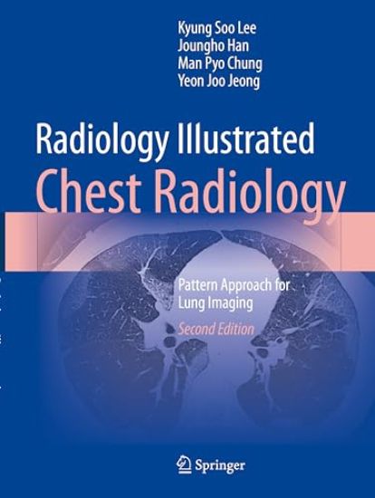
Radiology Illustrated: Chest Radiology

Radiology Illustrated: Chest Radiology

Kyung Soo Lee; Joungho Han; Man Pyo Chung; Yeon Joo Jeong

SPRINGER VERLAG, SINGAPORE
2025
nidottu
The purpose of this atlas is to illustrate how to achieve reliable diagnoses when confronted by the different abnormalities, or “disease patterns”, that may be visualized on CT scans of the chest. The task of pattern recognition has been greatly facilitated by the advent of high-technology CT such as helical and multidetector CT (MDCT) and dual-energy CT (DECT), and the focus of the book is very much on the role of state-of-the-art MDCT. A wide range of disease patterns and distributions are covered, with emphasis on the typical imaging characteristics of the various focal and diffuse lung diseases. In addition, clinical information relevant to differential diagnosis is provided and the underlying gross and microscopic pathology is depicted, permitting CT–pathology correlation. The entire information relevant to each disease pattern is also tabulated for ease of reference. This book will be an invaluable handy tool that will enable the reader to quickly and easily reach a diagnosis appropriate to the pattern of lung abnormality identified on CT scans. And in this second edition, some more signs and several new diseases and their pattern have been developed and updated. They will be provided with imaging, clinical relevance and CT-pathology correlation. Moreover, the chapters and disease patterns in their order of presentation shall be somewhat changed for readers’ easy legibility and understanding.
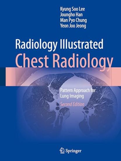
Radiology Illustrated: Chest Radiology

Radiology Illustrated: Chest Radiology

Kyung Soo Lee; Joungho Han; Man Pyo Chung; Yeon Joo Jeong

SPRINGER VERLAG, SINGAPORE
2024
sidottu
The purpose of this atlas is to illustrate how to achieve reliable diagnoses when confronted by the different abnormalities, or “disease patterns”, that may be visualized on CT scans of the chest. The task of pattern recognition has been greatly facilitated by the advent of high-technology CT such as helical and multidetector CT (MDCT) and dual-energy CT (DECT), and the focus of the book is very much on the role of state-of-the-art MDCT. A wide range of disease patterns and distributions are covered, with emphasis on the typical imaging characteristics of the various focal and diffuse lung diseases. In addition, clinical information relevant to differential diagnosis is provided and the underlying gross and microscopic pathology is depicted, permitting CT–pathology correlation. The entire information relevant to each disease pattern is also tabulated for ease of reference. This book will be an invaluable handy tool that will enable the reader to quickly and easily reach a diagnosis appropriate to the pattern of lung abnormality identified on CT scans. And in this second edition, some more signs and several new diseases and their pattern have been developed and updated. They will be provided with imaging, clinical relevance and CT-pathology correlation. Moreover, the chapters and disease patterns in their order of presentation shall be somewhat changed for readers’ easy legibility and understanding.

Radiology Illustrated: Chest Radiology

Radiology Illustrated: Chest Radiology

Kyung Soo Lee; Joungho Han; Man Pyo Chung; Yeon Joo Jeong

Springer-Verlag Berlin and Heidelberg GmbH Co. K
2016
nidottu
The purpose of this atlas is to illustrate how to achieve reliable diagnoses when confronted by the different abnormalities, or “disease patterns”, that may be visualized on CT scans of the chest. The task of pattern recognition has been greatly facilitated by the advent of multidetector CT (MDCT), and the focus of the book is very much on the role of state-of-the-art MDCT. A wide range of disease patterns and distributions are covered, with emphasis on the typical imaging characteristics of the various focal and diffuse lung diseases. In addition, clinical information relevant to differential diagnosis is provided and the underlying gross and microscopic pathology is depicted, permitting CT–pathology correlation. The entire information relevant to each disease pattern is also tabulated for ease of reference. This book will be an invaluable handy tool that will enable the reader to quickly and easily reach a diagnosis appropriate to the pattern of lung abnormality identified on CT scans.?