Kirjojen hintavertailu – 12 859 826 kirjaa ja 27 kauppaa

Kirjailija

A.L. Baert

Kirjat ja teokset yhdessä paikassa: 66 kirjaa, julkaisuja vuosilta 1999–2016, suosituimpiin kuuluu Radiology of Blunt Trauma of the Chest. Vertaile teosten hintoja ja tarkista saatavuus suomalaisista kirjakaupoista.

Nimi esiintyy myös muodoissa: A. L. Baert, A.L: Baert

66 kirjaa

Kirjojen julkaisuvuodet: 1999–2016.

Radiology of Blunt Trauma of the Chest

Radiology of Blunt Trauma of the Chest

P. Schnyder; A.L. Baert; M. Wintermark

Springer-Verlag Berlin and Heidelberg GmbH Co. K
2012
nidottu
During recent years important progress in intensive medicine methods has fundamentally changed the approach to and the management of acute chest trauma, which in developed countries is frequently related to road accidents, the number of which is still rising. It is important for the radiologist to become fully acquainted with the correct inter­ pretation of findings on conventional radiographs as well as on CT. Appropriate and rapid therapeutic action in life-threatening situations will frequently result from the skillful inter­ pretation of radiological findings and from the full integration of the radiologist as a well­ qualified member of the medical and surgical team, responsible for the global management of the chest trauma patient. I am indebted to Prof. P. Schnyder and Dr. M. Wintermark for their excellent work in col­ lecting the case material, which originates mainly from the University Hospital of Lausanne. of all diagnostic They have been able to present a comprehensive and up-to-date overview and interventional radiological aspects related to chest trauma. of great interest to all hospital-based radiologists, I am convinced that this volume will be and also to thoracic surgeons or intensive care physicians dealing with chest trauma patients. It is my sincere wish that this volume meet the same success with clinicians as many other volumes in our series. Leuven ALBERT L. BAERT Preface Diagnostic imaging has experienced astonishing developments during the relatively short period of its existence. We are just entering the second century of the medical use of x-rays.
Interventional Magnetic Resonance Imaging

Interventional Magnetic Resonance Imaging

A.L. Baert; R.W. Günther; G.K. von Schulthess

Springer-Verlag Berlin and Heidelberg GmbH Co. K
2012
nidottu
One of the most amazing and spectacular developments in modern radiology has been the rapid growth and expansion of so-called interventional radiology, which can also be described as minimally invasive therapy guided by radiological imaging. Many applications of this method are now widely in use in different organs, particularly in the vascular system. Everybody is well aware of the shortcomings and drawbacks of the radiological modalities currently used for guiding minimally invasive procedures. Ultrasound, although it has the advantage of being absolutely harmless to the patient and the operator, cannot be used for many procedures because it does not provide the precise anatomical information needed for a safe performance of these procedures. Rontgen rays provide superb anatomical insight to guide delicate manipulations inside the human body, but as operations tend to become longer and more complicated, the radia­ tion dose for patients, as well as for operators, is becoming an increasing source of concern. It is therefore logical that we should explore the possibilities for interventional radiological procedures provided by the latest imaging modality -magnetic resonan­ ce imaging -taking advantage of the specific physical properties of this method and the absence of ionizing radiation. It soon became evident that this new approach represents a tremendous challenge involving the development of new hardware and software, new catheters and other material that can be used in a magnetic environment, etc.
Radiology of Blunt Trauma of the Chest

Radiology of Blunt Trauma of the Chest

P. Schnyder; M. Wintermark; A. L. Baert

Springer-Verlag Berlin and Heidelberg GmbH Co. K
2000
sidottu
This volume exhaustively reviews all aspects of the radiology of blunt chest trauma, as well as the epidemiology, etiology, pathophysiology, clinical features, and pathology of such trauma. The interest of the book lies above all in the richness of its illustrations, resulting from an image data file spanning more than 10 years. Most radiological patterns described in the literature as well as some new ones are comprehensively discussed and displayed. Moreover, findings of conventional radiology, CT, and sometimes ultrasonography and angiography are displayed simultaneously, allowing comparison between the different imaging modalities. An introductory chapter describes the prevalence, risk factors, and biomechanics of blunt chest trauma. Blunt traumas of each anatomical compartment are then systematically reviewed according to a constant, concise, and structured approach. The book should be a valuable tool both for radiologists and for intensivists and other physicians involved in trauma care.
Imaging of Kidney Cancer

Imaging of Kidney Cancer

A.L. Baert; M.A. Bosniak

Springer-Verlag Berlin and Heidelberg GmbH Co. K
2016
nidottu
This is one of the first books to deal specifically with diagnostic imaging of the entire spectrum of kidney cancers. Both new and conventional imaging modalities are fully considered. After an introductory chapter on the histopathological classification of kidney cancers, the advantages and disadvantages of the various imaging modalities used in the diagnosis and assessment of disease extension are documented. Subsequent chapters offer an exhaustive description of the radiological features of the different histological subtypes of kidney cancer, with radiological and histological illustrations and tables. The latest innovations in interventional and minimally invasive procedures are also well covered. The book benefits from carefully chosen and technically excellent images. Each of the 24 chapters is written by an internationally acclaimed expert, making this book the most current and complete treatment of the subject available. It should be of great interest to radiologists, oncologists, and urologists.
Ultrasound of the Musculoskeletal System

Ultrasound of the Musculoskeletal System

Stefano Bianchi; A.L. Baert; Carlo Martinoli

Springer-Verlag Berlin and Heidelberg GmbH Co. K
2016
nidottu
A comprehensive reference and practical guide on the technology and application of ultrasound to the musculoskeletal system. It is organized into two main sections. The first is devoted to general aspects, while the second provides a systematic overview of the applications of musculoskeletal ultrasound in different areas of the body. Ultrasound scans are correlated with drawings, photographs, images obtained using other modalities, and anatomic specimens. There is a generous complement of high-quality illustrations based on high-end equipment. This book will acquaint beginners with the basics of musculoskeletal ultrasound, while more advanced sonologists and sonographers will learn new skills, means of avoiding pitfalls, and ways of effectively relating the ultrasound study to the clinical background.
Vascular Embolotherapy

Vascular Embolotherapy

A.L. Baert

Springer-Verlag Berlin and Heidelberg GmbH Co. K
2014
nidottu
Endovascular embolization therapy has made remarkable progress during the past two decades, driven primarily by improvements in digital imaging, breakthroughs in microcatheter technology and coil design, and the development of new embolic materials. Initially employed in the management of musculoskeletal trauma, embolization therapy now has a wide range of applications. It permits effective non-operative management of major hepatic, splenic, and renal injuries that once posed a tremendous challenge to the trauma surgeon and has also become an integral facet of the modern oncology center. Further important indications include uterine fibroids, vascular malformations, visceral aneurysms and GI bleeding. This volume and volume 2 present the current state of the art in this exciting and growing field. High informational content is supplemented by detailed graphics, providing a comprehensive reference work for both experts and novices in the field.
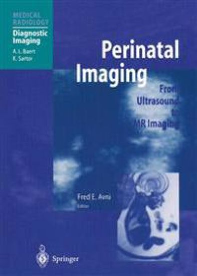
Perinatal Imaging

Perinatal Imaging

A.L. Baert

Springer-Verlag Berlin and Heidelberg GmbH Co. K
2014
nidottu
Fetal and perinatal medicine is a rapidly expanding field, and noninvasive imaging by means of ultrasonography and MRI is playing a major role in refining diagnosis and therapy. Recent technological advances in these imaging modalities now allow unprecedented morphological depiction of the fetus and excellent insight into complex pathologic conditions, as well as yielding superior guidance for therapeutic fetal inter­ ventions. I am very pleased that Professor F. Avni , a leading international pediatric radiologist, was prepared to take on the challenging task of preparing and editing this comprehen­ sive and up-to-date overview of our knowledge in the area of fetal and perinatal imaging. He has been successful in engaging well-known experts with outstanding qualifications in fetal imaging to join him in this venture. I would like to congratulate Professor Avni and all contributing authors most sincerely for their excellent work. I am confident that this outstanding volume will meet with great interest not only from general as well as specialized pediatric radiologists but also from neonatologists and pediatricians. I trust it will enjoy the same success as many previous volumes in this series. ALBERT L. BAERT Leuven Preface Fetal and perinatal medicine would not have developed without the extensive use of obstetric ultrasound (US). In order to be efficient, the examination has to be performed very carefully and by sonologists fully conversant with the normal and abnormal development of the fetus.
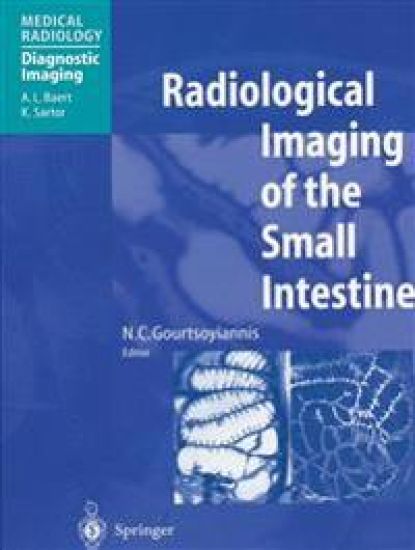
Radiological Imaging of the Small Intestine

Radiological Imaging of the Small Intestine

A.L. Baert

Springer-Verlag Berlin and Heidelberg GmbH Co. K
2014
nidottu
Whereas during the past decade endoscopy has become established as the leading means of diagnosis and management of diseases affecting the esophagus, starnach and large bowel, radiology has retained its pre-eminence for the clinical study and evaluation of the small bowel. This book provides unique coverage of all current radiological techniques used to study the small bowel, including not only barium studies and angiography but also cross-sectional methods such as uhrasound and computer tomography as well as nuclear medicine. Ernerging techniques such as magnetic resonance imaging are given close consideration, and interventional procedures are dealt with fully. Following an introduction on anatomy, physiology, and pathology pertinent to radiol­ ogy, the book describes in great detail common and less common congenital and development anomalies, trauma, infectious and infl.ammatory conditions, and tumors. The radiological accounts are illustrated by unique color photographs of pathological specimens. It is important for all professionals involved in the management of patients with small-bowel disease tobe critically informed about the specific advantages as well as the limitations of the various modern imaging techniques now employed for the in vivo morphological evaluation of the small bowel. This comprehensive book provides a timely update of our knowledge in this field and is a welcome addition to our series "Medical Radiology;' which aims to provide exhaustive coverage of modern diagnostic radiology. It will be of great interest for general and abdominal radiologists, gastroen­ terologists, and abdominal surgeons.
Recent Advances in Diagnostic Neuroradiology

Recent Advances in Diagnostic Neuroradiology

A.L. Baert

Springer-Verlag Berlin and Heidelberg GmbH Co. K
2014
nidottu
Diagnostic neuroradiology is undergoing such rapid change that standard texts are quickly becoming outdated in important respects. Recent Advances in Diagnostic Neuroradiology is designed to complement the general textbooks of neuroradiology by documenting and discussing the progress that has been achieved. Following six introductory chapters, 26 important topics in brain and spinal imaging are discussed in detail, with appropriate illustrations and a review of the most recent literature. Each of these topics has specifically been chosen in order to summarize recent developments and to document the state of the art in the field. This book, written by acknowledged experts in the field, will be of relevance and importance to all with an interest in neuroradiology.
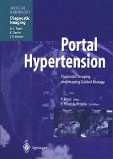
Portal Hypertension

Portal Hypertension

A.L. Baert

Springer-Verlag Berlin and Heidelberg GmbH Co. K
2013
nidottu
Portal hypertension is a relatively common condition, the complications of which are frequently life threatening. Patients with portal hypertension represent an important por­ tion of the daily clinical work of abdominal radiologists, gastroenterologists and abdominal surgeons. During the past two decades the development of new radiological devices and of new percutaneous image-guided treatment methods has resulted in fundamental changes in the clinical management of patients with portal hypertension. During the same period tremen­ dous progress was achieved in the visualization of the numerous morphological and func­ tional changes observed in patients with portal hypertension, due to the clinical introduc­ tion of the new cross-sectional imaging methods: ultrasonography, computed tomography and magnetic resonance imaging. I am very grateful to Prof. Plinio Rossi, an internationally recognized leader in the field of interventional radiology, for accepting the challenging task of compiling and presenting in a structured way the numerous facts and data that have become available during the past few years in this rapidly developing field. The editor has been inspired and successful in his selection of the impressive group of international experts that has contributed to many chapters of this volume.
Clinical and Radiological Aspects of Myopathies

Clinical and Radiological Aspects of Myopathies

J. A. L. Bulcke; A. L. Baert

Springer-Verlag Berlin and Heidelberg GmbH Co. K
2013
nidottu
One of the most puzzling and striking features of many of the genetically­ determined progressive neuromuscular diseases such as the spinal muscular atrophies and the muscular dystrophies is that muscular wasting and weak­ ness in these cases is curiously selective, at least in the early stages, pick­ ing out certain skeletal muscles and sparing others. The diagnosis of these conditions has largely depended in the past upon the recognition of specific patterns of involvement of individual muscles and muscle groups, taken along with information derived from the mode of inheritance within the in­ dividual family and the results of special investigations. The investigations of most value have proved to be serum enzyme studies, electromyography and related techniques, and muscle biopsy. The advent of CT scanning has, however, introduced a new dimension; as the authors of this interesting monograph have clearly demonstrated, it is now possible, using the whole­ body scanner, to define patterns of muscular atrophy in the limbs and trunk much more precisely than by any other method. Not only does this techni­ que demonstrate which muscles are involved, but the changes in relative density provide useful information about the severity of the process and about the progress of the disease if the studies are performed serially. This monograph is pleasantly written and most attractively illustrated.
Radiological Imaging of the Neonatal Chest

Radiological Imaging of the Neonatal Chest

A.L. Baert

Springer-Verlag Berlin and Heidelberg GmbH Co. K
2013
nidottu
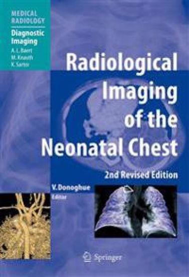
This second, revised edition of “Radiological Imaging of the Neonatal Chest” provides a comprehensive and up-to-date discussion of the subject. It is written primarily from the point of view of the paediatric radiologist but will be of particular interest to all antenatal ultrasonographers, neonatologists, paediatric cardiologists, paediatricians and paediatric surgeons. It includes an update on clinical management and appraises the advantages of the various techniques available to image the newborn chest. There is particular emphasis on the impact of recent therapeutic advances on imaging findings. Dedicated chapters are included on antenatal and postnatal imaging of chest malformations, upper airway problems, infection and congenital heart disease, with special emphasis on the current role of magnetic resonance imaging, computed tomography and interventional therapy. This well-illustrated book contains important information for all those involved in caring for the neonate.
Transfontanellar Doppler Imaging in Neonates

Transfontanellar Doppler Imaging in Neonates

A. Couture; A.L. Baert; C. Veyrac

Springer-Verlag Berlin and Heidelberg GmbH Co. K
2012
nidottu
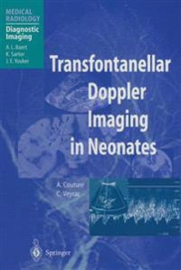
Transfontanellar Doppler imaging was until recently an emerging new diagnostic modality that was not widely used in clinical practice. Dr. A. Couture and Dr. C. Veyrac have pioneered and incessantly developed the tech­ and neonatal brain scanning until it has now been matured into an nique of prenatal indispensable tool for the correct management of infants. Building upon the technical progress in the manufacturing of scanning probes and ultrasonic equipment, the authors have been able to accumulate a unique and exceptional body of knowledge on fetal and neonatal brain scanning. Their book provides exquisite information not only on the normal neonatal brain, but also on the various pathological conditions and malformations involving the fetal and neonatal brain as provided by ultrasound examination. The book contains numerous illustrative examples, selected from the outstanding personal files of the authors. Transfontanellar Doppler imaging, because of its great impact on the management of neonates suspected of brain abnormalities, should be available to all pediatric and general radiologists involved in brain scanning in the pediatric age group. They, and also neurologists and neurosurgeons, will find this volume a unique source of information and knowledge. I congratulate the editors on their outstanding performance and I wish this volume great success within the medical community. Leuven ALBERT L. BAERT Preface Alain Couture belongs to the breed of pioneers. This requires several unusual traits: orginality, tenacity and vision, and of course hard work.
Liver Malignancies

Liver Malignancies

A.L. Baert

Springer-Verlag Berlin and Heidelberg GmbH Co. K
2012
nidottu
A comprehensive and up-to-date overview of the role of diagnostic and interventional radiology in respect of liver malignancies. Following background chapters on anatomy, epidemiology, and clinicopathologic features, each of the diagnostic imaging techniques is carefully appraised, focusing on new developments in equipment and contrast agents. The interventional therapeutic approaches to primary and secondary hepatic malignancies are then discussed in depth, as well as such special topics as liver tumors in children and hepatic transplantation. Written by leading experts from around the world, this will prove to be an indispensable source of information for both clinicians and researchers.
Radiology of Peripheral Vascular Diseases

Radiology of Peripheral Vascular Diseases

A.L. Baert

Springer-Verlag Berlin and Heidelberg GmbH Co. K
2012
nidottu
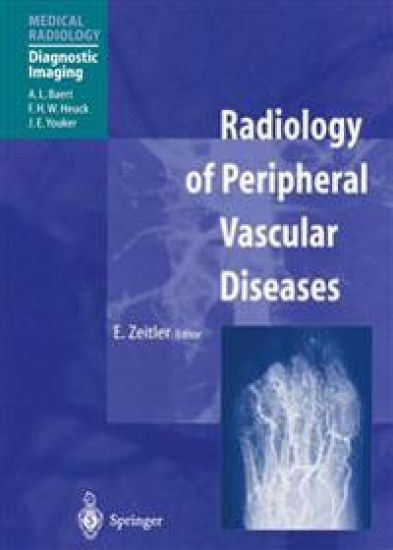
Over the last decade, important advances have been made in the radiological man­ agement of peripheral vascular diseases. In the diagnostic field, the role of con­ ventional angiography has diminished considerably in favor of new, non-invasive diagnostic modalities such as color Doppler imaging and magnetic resonance angiography. Percutaneous radiological treatment of arterial diseases of the limbs has revolu­ tionized therapeutic concepts in this area. Professor E. Zeitler is a renowned international expert in the field of peripheral He was one of the pioneers in investigating and developing per­ vascular diseases. cutaneous catheter treatment of arterial stenosis of peripheral arteries. This volume is based upon his unique lifetime's experience of treating patients presenting with peripheral vascular diseases. Moreover, Professor Zeitler was very successful in engaging a group of excellent authors, all specialists in their field, to contribute individual chapters. Together, they have suceeded in producing one of the most comprehensive books on the modern radiological approach to patients with peripheral vascular diseases. It is my firm belief that this volume will be of great interest to radiologists, angiologists, and vascular surgeons alike by providing them with the latest knowl­ edge and information in the field of radiology. I wish this volume the same success as the many other volumes already published in the book series "Medical Radiology." Any constructive criticism that the reader may wish to affer is welcomed.
Medical Imaging of the Spleen

Medical Imaging of the Spleen

A.L. Baert

Springer-Verlag Berlin and Heidelberg GmbH Co. K
2012
nidottu
I would like to express my great appreciation to the editors of this volume, Prof. A. De Schepper and Dr. F. Vanhoenacker, for all their efforts and their enthusiastic commitment to this book. I am very grateful to them for managing to involve so many leading experts, resulting in a much -needed high -quality and absolutely up-to-date text­ book on the spleen, an organ which has not always received the attention it deserves from radiologists. As series editor, I am particularly grateful to the volume editors for adhering strictly to the planed schedule. Their punctuality enabled Springer-Verlag to reduce the pro­ duction time of this volume to a minimum. I am very much convinced that this volume on the spleen responds to an important need within the radiological community and that many radiologists will greatly bene­ fit from it. However, surgeons and internal medicine specialists will also be interested in this excellent comprehensive overview of modern radiological imaging of splenic dis­ orders. Leuven ALBERT L. BAERT Preface Not infrequently, the spleen is regarded as the "silent and forgotten" organ of the abdomen. Though primary splenic diseases are rare, the spleen is a frequent site of sec­ ondary manifestations in a wide range of hematological, immunological, oncological, infectious, vascular and systemic disorders. Despite this broad spectrum of splenic dis­ eases, radiological literature on splenic pathology is rather sparse.
Radiology of AIDS

Radiology of AIDS

A.L. Baert

Springer-Verlag Berlin and Heidelberg GmbH Co. K
2012
nidottu
Although astonishing progress has been achieved during recent years in the treatment of patients afflicted with AIDS, this disease is still spreading in much of the developing world, where economic resources are limited. Physicians working in these geographic areas in­ creasingly face the dramatic and devastating consequences of this epidemic and will cer­ tainly benefit from the information imparted in this book. Indeed, it has now been well established that radiologic studies playa key role in the diagnosis and management of patients with HIV infection. In the Western world the positive effects of new potent drugs are already discernible, resulting in decreased mortality from the illness. But as AIDS patients survive longer, the medical profession is confronted with changing radiologic findings related to longstanding AIDS. This superb book provides a very comprehensive overview of our current knowledge about radiologic imaging features in AIDS. I would like to congratulate most sincerely the Editors for their tremendous field work, their enthusiasm, and their high level of expertise, which have resulted in the publication of this outstanding volume. To assist them in their task, they have recruited numerous leading specialists in the field. Meticulous editing has reduced the repetition that otherwise can easily occur in a multiauthored textbook. I am convinced that this comprehensive and richly illustrated work will be of great interest to all clinical disciplines involved in the management of AIDS patients.
CT of the Peritoneum

CT of the Peritoneum

Armando Rossi; A.E. Cardinale; Giorgio Rossi; A.L. Baert

Springer-Verlag Berlin and Heidelberg GmbH Co. K
2012
nidottu
I have not embarked on the foreword to this scientific monograph by Armando and Gior­ gio ROSSI in the expectation that it will be an easy task, because these two authors are the last remaining members of a family that has left its mark in the field of radiology in our country: therefore, the writer's enthusiasm and detachment could be jeopardized by mem­ ories of his own teachers and elders and the respect he still feels towards them. The line stretches from Armando Rossi Sr., a pioneer in the field of radiology in Italy, a scientist and a versatile teacher, a student of Beclere and Busi, to Lucio Rossi, an eminent teacher,a learned man and a gentleman. An official biographyofArmando Rossi shows that in his last years, his wide didactic interests were directed towards his own family, leading him to devote his attention to those of his grandchildren who were then getting ready to embrace the medical profession.
Frontiers in European Radiology

Frontiers in European Radiology

A. L. Baert; F. H. W. Heuck

Springer-Verlag Berlin and Heidelberg GmbH Co. K
2011
nidottu
Contents Abdominal and Thoracic Magnetic Resonance Angiography: Challenges, Technical Approaches and First Clinical Applications. H. U. Kauczor, R. Hausmann, W. Semmler Magnetic Resonance Spectroscopy of Skeletal Muscle. F. Tr{ber, W. A. Kaiser, G. Layer,C. Kuhl, M. Reiser Hepatic Tumors: Relaxometry and Quantitative Tissue Characterization with Magnetic Resonance Imaging. A. Steudel, F. Tr{ber, M. Reiser Liver-Specific Particulate Contrast Agents: An Overview I. Ivancev, A. Lunderquist Contrast Medium Induced Nephropathy:Animal Experiments H. S. Thomsen et al. Rheological Properties of Blood after Diagnostic Cardiac Catheterisation with Iopromide. R. Bach et al. Color Coded Doppler Sonography of the Male Urethra During Voiding: New Observations of Flow in an Aqueous Medium and Their Physical Foundations. P. H}bsch, F. Hager, E. Benes Development of a Model of Arterial Stenosis Analysis Using Digital Subtraction Angiography: A Parametric Study of Image Quality. C. Renaudin, I. Magnin, A. Amiel Arterial Calcifications in Thoracoabdominal Computer Tomography: Frequency and Correlation with Risk Factors and Cardiovascular Diseases. J. G|rich et al.