Kirjojen hintavertailu – 12 859 826 kirjaa ja 27 kauppaa
Kirjailija
A.L. Baert
Kirjat ja teokset yhdessä paikassa: 66 kirjaa, julkaisuja vuosilta 1999–2016, suosituimpiin kuuluu Radiology of Blunt Trauma of the Chest. Vertaile teosten hintoja ja tarkista saatavuus suomalaisista kirjakaupoista.
Nimi esiintyy myös muodoissa: A. L. Baert, A.L: Baert
This book, edited by Prof. PLINIO ROSSI, an internationally well-known expert in biliary tract radiology, provides in 31 chapters a very comprehensive update on both diagnostic and therapeutic radiology of the biliary tract. First, a very complete and interesting overview of the normal morphology of the ana tomical area is presented by an anatomist, a radiologist, and a surgeon. This is followed by a discussion not only of the well-known and accepted imaging modalities, but also of the newer ones such as, magnetic resonance imaging and endoluminal and laparoscopic ultra sound. The book thus illustrates very nicely the diversity of diagnostic radiological tech niques that are currently available. Considerable technical progress and remarkable new insights in biliary tract radiology have been achieved in the field of the radiological interventional approach during recent years. Therefore, much emphasis has been placed in this book on the therapeutic possibili ties of the radiologist using the percutaneous minimally invasive approach for the treat ment of the various pathological conditions of the biliary tract. It is most impressive to note the substantial progress in terms of patient comfort achieved by using the percutaneous as compared to the surgical methods. These new methods also result in shorter hospitalization times and thereby help to reduce health care costs. This book provides the interested reader with a fascinating and very complete over view of these new procedures.
Few fields have witnessed such impressive advances as the application of computer technology to radiology. The progress achieved has revolutionized diagnosis and greatly facilitated treatment selection and accurate planning of procedures. This book, written by leading experts from many different countries, provides a comprehensive and up-to-date overview of the role of 3D image processing. The first section covers a wide range of technical aspects in an informative way. This is followed by the main section, in which the principal clinical applications are described and discussed in depth. To complete the picture, the final section focuses on recent developments in functional imaging and computer-aided surgery. This book will prove invaluable to all who have an interest in this complex but vitally important field.
Sports medicine is acquiring an important role owing to the increasing number of sports-active people and professional athletes. Accurate diagnosis of the different pathological conditions is therefore of fundamental importance. This book provides an overview of the most frequently observed conditions and correlates them with sports activities, as well as documenting relatively unknown lesions of increasing significance. Diagnostic techniques are described and compared, and their roles defined; interpretative pitfalls are highlighted. All of the contributing authors have distinguished themselves in the field and have a deep knowledge of the problem involved in the diagnosis and classification of sports injuries.
Imaging studies are playing an increasingly role in the evaluation of endocrine diseases; accordingly, familiarity with the specific indications for the various modalities, and with the characteristic findings, is essential. This multi-author work, which is intended for both radiologists and endocrinologists, considers the role of all the recent imaging techniques, including ultrasound (particular color Doppler), computed tomography, MRI, and scintigraphy. Following an extensive introduction on the pituitary, subsequent chapters discuss in detail the normal anatomy and pathology of the female and male reproductive systems. Remaining chapters provide state-of-the-art data on the thyroid, parathyroids, pancreatic endocrine tumors, adrenal glands, hormonal tumors (carcinoids and MEN), and imaging of the complications of hormone therapy.
The use 0\ contrast media within diagnostic radiology has been firmly established for many years now. Radiologic contrast media render immense services in daily clinical practice to innumerable patients all over the world. When the new cross sectional modalities such as computed tomography and magnetic resonance ima ging were introduced into clinical practice it rapidly became evident that the judi cious use of contrast media allows the optimal exploitation of the great diagnostic potential of these revolutionary methods. More recently, efforts to create specific contrast media for ultrasound imaging have been successful, and the first benefi cial results of their application in daily practice are now evident. The aim of this book is to bring together the results of the tremendous research that has been performed during recent years in order to develop specific contrast media for magnetic resonance imaging and ultrasonography and also to develop safer contrast media for the classical radiologic techniques as well as computed tomography. Moreover, the book provides a comprehensive overview of the indica tions for and applications of these new contrast media in clinical radiology. Finally, risks, side-effects, and complications which may occur with the use of these drugs are covered appropriately. The three editors are leading and well-known experts in their specific field. They have been very successful in ensuring the collaboration of so many intern~tionally known specialists to cover the different topics dealt with in this book.
This is a comprehensive textbook on imaging of the bony pelvis and hip joint that provides a detailed description of the techniques and imaging findings relevant to this complex anatomical region. In the first part of the book, the various techniques and procedures employed for imaging the pelvis and hip are discussed in detail. The second part of the book documents the application of these techniques to the diverse clinical problems and diseases encountered. Among the many topics addressed are congenital and developmental disorders including developmental dysplasia of the hip, irritable hip and septic arthritis, Perthes' disease and avascular necrosis, slipped upper femoral epiphysis, bony and soft tissue trauma, arthritis, tumours and hip prostheses. Each chapter is written by an acknowledged expert in the field, and a wealth of illustrative material is included. This book will be of great value to musculoskeletal and general radiologists, orthopaedic surgeons and rheumatologists.
At last there is on the market a comprehensive reference and practical guide on the application of US to penile diseases and conditions. This is quite simply the most extensive textbook on the subject. After introductory chapters on technical requirements and penile anatomy, subsequent chapters offer a systematic overview of the diverse applications of color Doppler US.
1 Introduction Christian Schiepers went major revisions. A few were updated and had CONTENTS only minor revisions (Chaps. 4, 7 and 15,) and two 1. 1 Perspective 1 were left unchanged and re-printed from the first 1. 2 Objectives 2 edition. Our selection is aimed at elucidating key 1. 3 Clinical Overview 2 processes in cellular mechanisms of the human body, 1. 4 Basics of Diagnostic Nuclear Medicine 4 under normal conditions as well as in disease. 1. 5 Future Perspective 4 In the present revised volume of Diagnostic Nuclear 1. 1 Medicine, the advancements in the field of nuclear Perspective medicine (NM) are presented with an emphasis on progress in the beginning of this millennium. The NM started as a field where radioactive products name ‘molecular imaging’ is used more frequently were put to use for the benefit of mankind, e. g. t- for diagnostic NM imaging, but is not commonplace. roid scintigraphy and therapy. The performed stud- We will use the traditional term NM. The various ies in the field have fluctuated tremendously since contributions in this imaging field such as new trac- those early years. Flow imaging of the brain was a ers and equipment, modifications of existing tests, frequent procedure in the NM clinic until CT was diagnostic algorithms, and general applications for introduced. Later on, sophisticated triggering te- whole body imaging are discussed.
Since the first edition of this book, sonography of the peripheral nervous system has evolved further. Not only has knowledge of the sonographic features of common neurological disorders deepened, but constant research has led to the application of sonography in the diagnosis and guided treatment of disorders that were not previously accessible. This second, revised edition reflects these ongoing developments in various ways: many state of the art high-resolution images are included, the text has been adapted to reflect the current state of the literature, and information is presented using a more modern layout. Anatomic/sonographic correlation is considered in depth, and examination technique with modern sonography equipment is explained. This book provides a practical, clinically oriented overview of all aspects of sonographic diagnosis and interventional therapy of the peripheral nervous system and will be of value to all with an interest in this field.
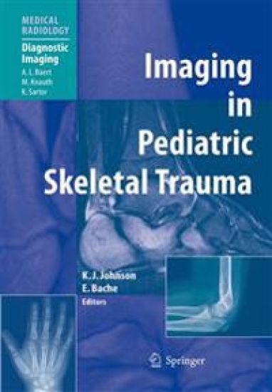
This is a comprehensive textbook on imaging of pediatric skeletal trauma. It provides a detailed description of the techniques used and the imaging findings, detailing their clinical relevance. In the first part of the book the various techniques and procedures needed to successfully image both acute and more chronic skeletal injuries are discussed in depth. Individual chapters are devoted to radiography, CT, MRI, ultrasound, and nuclear medicine. The second part of the book documents the different types of skeletal injury, and the chapters are arranged according to anatomical site. In addition, chapters are included on growth plate injuries, non-accidental injuries, and normal variants that can imitate fractures. Each chapter is written by an expert in the field and a wealth of illustrative material is included. This book is designed to be of value to all radiologists who are involved with pediatric trauma, whether trainees or consultants, and also to orthopedic surgeons.
my fellows and residents. They have missed their teacher quite a bit during the “hot” periods of this project, and they paid me back, not by compla- ing, but by asking pertinent questions and by providing me with interesting cases. I would like to thank Peter Jaksch and Walter Klepetko from the Lung Transplantation Unit of my home University of Vienna, Austria. Their clinical and surgical competence, combined with their reliability, have made our daily collaboration a productive and enjoyable partnership. I owe my deep gratitude to Pierre Alain Gevenois, Department of Radiology, and Marc Estenne, Lung Transplantation Unit, both at the Hôpital Erasme, University of Brussels, B- gium. Their support, expertise, and friendship have aided our fruitful research collaboration in the ? eld of lung transplantation over the past decade. Without their ongoing input, many things would simply not have happened. I would also like to thank Christiane Knoop, Alain Van Muylem, and Denis Tack, all from the same institution, for satisfying my often intrusive (and most likely annoying) avidity for their help and knowledge. It is to Ursula N. Davis at Springer-Verlag that I sincerely apologize for my sometimes more than undulating working rhythm, a rhythm she tolerated with admirable patience and humor. Without her moral support and her enduring determination, I would not have made it through this project. I am also grateful to Prof. Albert L.
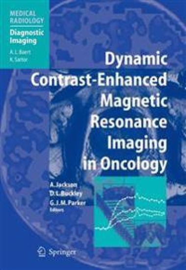
Dynamic contrast-enhanced MRI is now established as the methodology of choice for the assessment of tumor microcirculation in vivo. This is assisting clinical practitioners in the management of patients with solid tumors and is finding prominence in the assessment of tumor treatments, including anti-angiogenics, chemotherapy, and radiotherapy. In this book, targeted at both clinical practitioners and basic scientists, the principles of the methods, their practical implementation, and their application to specific tumor types are discussed by the leading authorities in the field today. The book will serve as an invaluable single-volume reference covering all the latest developments in contrast-enhanced oncological MRI.
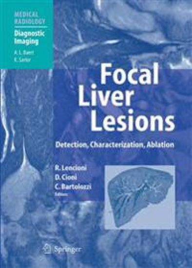
Few fields of medicine have witnessed such impressive progress as the diagnosis and treatment of liver tumors. Advances in imaging technology, the development of novel contrast agents, and the introduction of optimized scanning protocols have greatly facilitated the non-invasive detection and characterization of focal liver lesions. Furthermore, image-guided techniques for percutaneous tumor ablation have become an accepted alternative treatment for patients with inoperable liver cancer. This book provides a comprehensive and up-to-date overview of the role of diagnostic and interventional radiology in respect of liver tumors. The volume moves from background sections on methodology and segmental liver anatomy to the main sections on the diagnosis of benign and malignant liver lesions. An integrated approach, focused on the correlation of ultrasound, CT, and MR imaging findings, is presented. Finally, a full section describes the principles, methods, and results of percutaneous tumor ablation techniques.
Multidetector-row CT has dramatically improved the results of computed tomography in all clinical applications, but its beneficial impact has been most striking in vascular imaging. The simplicity of acquisition and the wide availability of equipment make this modality especially suitable for routine clinical application. In this book the basic aspects of multidetector-row CT angiography are comprehensively reviewed. Individual chapters are included on technical principles, image processing techniques and contrast agent administration. All clinical applications are then discussed in depth, with lucid descriptions of the examination technique for particular clinical indications and of the findings that characterize specific diseases. Limitations and advantages in comparison with other imaging modalities are considered. A large number of high-quality black and white and color illustrations help to explain the clinical findings.
Te idea of publishing a book came into my mind to the description of the imaging fndings of parasitic di- s (M. C. H. ) when a few years ago I submitted a manuscript eases only. Parasitic diseases are not confned only to the entitled “Imaging of Parasitic Diseases of the Hepato - tropical and subtropical areas; indeed, they are increasing biliary System” for publication in the major prestigious worldwide because of travel, immunosuppression, and radiology journals. Te manuscript was rejected, but an other behavioral and living defciencies, especially poor anonymous reviewer commented that the manuscript hygiene and sanitation. Some of these diseases are prev- would be more suitable for publication as a chapter in a lent or clustered in poor or developing countries, but the book. I took up the challenge and began the search for majority are encountered worldwide, even in industri- co-editors and authors to write other chapters. At this ized and developed countries. point, I would like to thank the reviewer for giving me In the radiologic bibliography, there is a shortage of the idea and throwing down the challenge. books on the subject of “Imaging of Infection. ” Tis book Te aim of this book is to introduce the concept of is intended to partly rectify this shortfall and is aimed at “Imaging of Infection,” which includes bacterial, viral, radiologists all over the world. fungal, and parasitic infections.
MRI and CT exquisitely depict the anatomy of the female pelvis and offer fascinating diagnostic possibilities in women with pelvic disorders. This volume provides a comprehensive account of the use of these cross-sectional imaging techniques to identify and characterize developmental anomalies and acquired diseases of the female genital tract. Both benign and malignant diseases are considered in depth, and detailed attention is also paid to normal anatomical findings and variants. Further individual chapters focus on the patient with pelvic pain and the use of MRI for pelvimetry during pregnancy and the evaluation of fertility. Throughout, emphasis is placed on the most recent diagnostic and technical advances, and the text is complemented by many detailed and informative illustrations. All of the authors are acknowledged experts in diagnostic imaging of the female pelvis, and the volume will prove an invaluable aid to everyone with an interest in this field.
Few fields have witnessed such impressive advances as image processing in radiology. The progress achieved has revolutionized diagnosis and greatly facilitated treatment selection and accurate planning of procedures. This book, written by leading experts from many countries, provides a comprehensive and up-to-date description of how to use 2D and 3D processing tools in clinical radiology. The first section covers a wide range of technical aspects in an informative way. This is followed by the main section, in which the principal clinical applications are described and discussed in depth. To complete the picture, a third section focuses on various special topics. The book will be invaluable to radiologists of any subspecialty who work with CT and MRI and would like to exploit the advantages of image processing techniques. It also addresses the needs of radiographers who cooperate with clinical radiologists and should improve their ability to generate the appropriate 2D and 3D processing.
Continuous technical developments of radiological imaging techniques have not only improved the potential of organ-based radiological diagnostics, but now also allow for dedicated whole-body exams which may be used in the field of screening and preventive diagnosis. The concept of this book is to provide clinicians with a broader understanding of screening and preventive diagnosis using radiological imaging. The first part of the book is dedicated to the fundamentals of screening and preventive diagnosis, and will comprise chapters on epidemiology and pathology, technical and organizational aspects of radiological screening, and chapters on legal issues and cost-risk analysis. The second part of the book will discuss the most important practical examples of radiological screening and surveillance, both for unselected populations, as well as for individual risk groups.
This second edition of "Radiology of Osteoporosis" has been fully updated so as to represent the current state of the art. It provides a comprehensive overview of osteoporosis, the pathologic conditions that give rise to osteoporosis, and the complications that are frequently encountered. After initial chapters devoted to pathophysiology, the presentation of osteoporosis on conventional radiographs is illustrated and discussed. Thereafter, detailed consideration is given to each of the measurement methods employed to evaluate osteoporosis, including dual x-ray absorptiometry, vertebral morphometry, spinal and peripheral quantitative computed tomography, quantitative ultrasound, and magnetic resonance imaging. The role of densitometry in daily clinical practice is appraised. Finally, a collection of difficult cases involving pitfalls is presented, with guidance to their solution. The information contained in this volume will be invaluable to all with an interest in osteoporosis.